| Host: |
Rabbit |
| Applications: |
WB/IHC/IF |
| Reactivity: |
Human/Mouse/Rat |
| Note: |
STRICTLY FOR FURTHER SCIENTIFIC RESEARCH USE ONLY (RUO). MUST NOT TO BE USED IN DIAGNOSTIC OR THERAPEUTIC APPLICATIONS. |
| Short Description: |
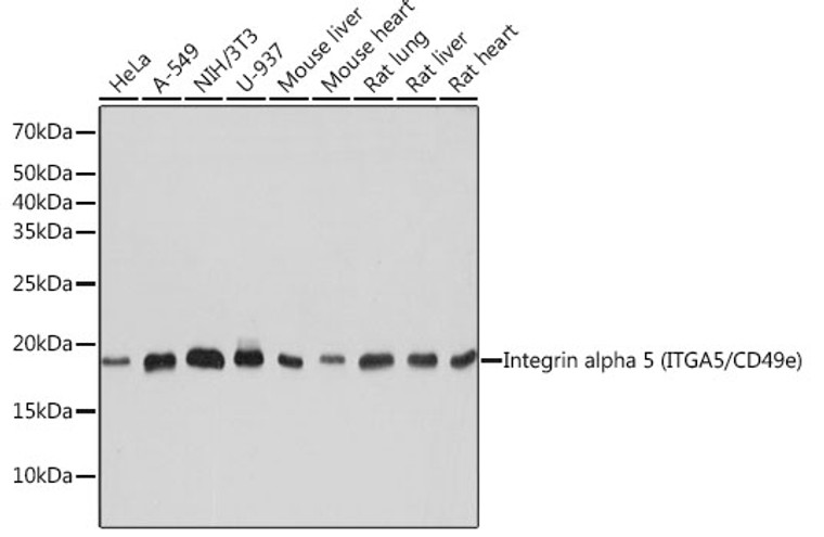
Rabbit monoclonal antibody anti-Integrin alpha 5 (950-1049) is suitable for use in Western Blot, Immunohistochemistry and Immunofluorescence research applications. |
| Clonality: |
Monoclonal |
| Clone ID: |
S6MR |
| Conjugation: |
Unconjugated |
| Isotype: |
IgG |
| Formulation: |
PBS with 0.02% Sodium Azide, 0.05% BSA, 50% Glycerol, pH7.3. |
| Purification: |
Affinity purification |
| Dilution Range: |
WB 1:500-1:2000IHC-P 1:50-1:200IF/ICC 1:50-1:200FC (Intra) 1:50-1:200 |
| Storage Instruction: |
Store at-20°C for up to 1 year from the date of receipt, and avoid repeat freeze-thaw cycles. |
| Gene Symbol: |
ITGA5 |
| Gene ID: |
3678 |
| Uniprot ID: |
ITA5_HUMAN |
| Immunogen Region: |
950-1049 |
| Immunogen: |
A synthetic peptide corresponding to a sequence within amino acids 950-1049 of human Integrin alpha 5 (ITGA5/CD49e) (P08648). |
| Immunogen Sequence: |
HQPFSLQCEAVYKALKMPYR ILPRQLPQKERQVATAVQWT KAEGSYGVPLWIIILAILFG LLLLGLLIYILYKLGFFKRS LPYGTAMEKAQLKPPATSDA |
| Tissue Specificity | Expressed in placenta (at protein level). |
| Post Translational Modifications | Proteolytic cleavage by PCSK5 mediates activation of the precursor. |
| Function | Integrin alpha-5/beta-1 (ITGA5:ITGB1) is a receptor for fibronectin and fibrinogen. It recognizes the sequence R-G-D in its ligands. ITGA5:ITGB1 binds to PLA2G2A via a site (site 2) which is distinct from the classical ligand-binding site (site 1) and this induces integrin conformational changes and enhanced ligand binding to site 1. ITGA5:ITGB1 acts as a receptor for fibrillin-1 (FBN1) and mediates R-G-D-dependent cell adhesion to FBN1. ITGA5:ITGB1 acts as a receptor for fibronectin (FN1) and mediates R-G-D-dependent cell adhesion to FN1. ITGA5:ITGB1 is a receptor for IL1B and binding is essential for IL1B signaling. ITGA5:ITGB3 is a receptor for soluble CD40LG and is required for CD40/CD40LG signaling. (Microbial infection) Integrin ITGA5:ITGB1 acts as a receptor for Human metapneumovirus. (Microbial infection) Integrin ITGA2:ITGB1 acts as a receptor for Human parvovirus B19. (Microbial infection) In case of HIV-1 infection, the interaction with extracellular viral Tat protein seems to enhance angiogenesis in Kaposi's sarcoma lesions. |
| Protein Name | Integrin Alpha-5Cd49 Antigen-Like Family Member EFibronectin Receptor Subunit AlphaIntegrin Alpha-FVla-5Cd Antigen Cd49e Cleaved Into - Integrin Alpha-5 Heavy Chain - Integrin Alpha-5 Light Chain |
| Database Links | Reactome: R-HSA-1566948Reactome: R-HSA-1566977Reactome: R-HSA-202733Reactome: R-HSA-216083Reactome: R-HSA-445144Reactome: R-HSA-8941332Reactome: R-HSA-9634597 |
| Cellular Localisation | Cell MembraneSingle-Pass Type I Membrane ProteinCell JunctionFocal Adhesion |
| Alternative Antibody Names | Anti-Integrin Alpha-5 antibodyAnti-Cd49 Antigen-Like Family Member E antibodyAnti-Fibronectin Receptor Subunit Alpha antibodyAnti-Integrin Alpha-F antibodyAnti-Vla-5 antibodyAnti-Cd Antigen Cd49e Cleaved Into - Integrin Alpha-5 Heavy Chain - Integrin Alpha-5 Light Chain antibodyAnti-ITGA5 antibodyAnti-FNRA antibody |
Information sourced from Uniprot.org
12 months for antibodies. 6 months for ELISA Kits. Please see website T&Cs for further guidance


