| Host: |
Rabbit |
| Applications: |
WB |
| Reactivity: |
Human/Mouse/Rat |
| Note: |
STRICTLY FOR FURTHER SCIENTIFIC RESEARCH USE ONLY (RUO). MUST NOT TO BE USED IN DIAGNOSTIC OR THERAPEUTIC APPLICATIONS. |
| Short Description: |
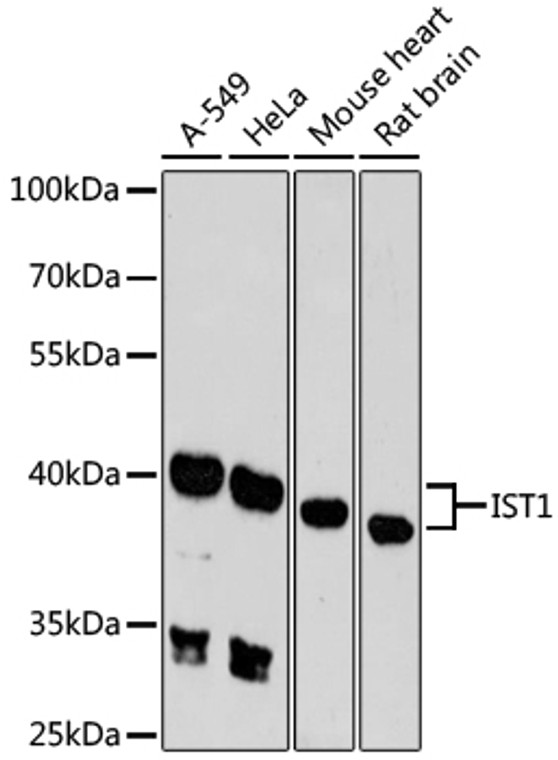
Rabbit polyclonal antibody anti-IST1 (1-335) is suitable for use in Western Blot research applications. |
| Clonality: |
Polyclonal |
| Conjugation: |
Unconjugated |
| Isotype: |
IgG |
| Formulation: |
PBS with 0.01% Thimerosal, 50% Glycerol, pH7.3. |
| Purification: |
Affinity purification |
| Dilution Range: |
WB 1:500-1:2000 |
| Storage Instruction: |
Store at-20°C for up to 1 year from the date of receipt, and avoid repeat freeze-thaw cycles. |
| Gene Symbol: |
IST1 |
| Gene ID: |
9798 |
| Uniprot ID: |
IST1_HUMAN |
| Immunogen Region: |
1-335 |
| Immunogen: |
Recombinant fusion protein containing a sequence corresponding to amino acids 1-335 of human IST1 (NP_001257906.1). |
| Immunogen Sequence: |
MLGSGFKAERLRVNLRLVIN RLKLLEKKKTELAQKARKEI ADYLAAGKDERARIRVEHII REDYLVEAMEILELYCDLLL ARFGLIQSMKELDSGLAESV STLIWAAPRLQSEVAELKIV ADQLCAKYSKEYGKLCRTNQ IGTVNDRLMHKLSVEAPPKI LVERYLIEIAKNYNVPYEPD SVVMAEAPPGVETDLIDVGF TDDVKKGGPGRGGSGGFTAP VGGPDGTVPMPMPMPMPMP |
| Function | ESCRT-III-like protein involved in cytokinesis, nuclear envelope reassembly and endosomal tubulation. Is required for efficient abscission during cytokinesis. Involved in recruiting VPS4A and/or VPS4B to the midbody of dividing cells. During late anaphase, involved in nuclear envelope reassembly and mitotic spindle disassembly together with the ESCRT-III complex: IST1 acts by mediating the recruitment of SPAST to the nuclear membrane, leading to microtubule severing. Recruited to the reforming nuclear envelope (NE) during anaphase by LEMD2. Regulates early endosomal tubulation together with the ESCRT-III complex by mediating the recruitment of SPAST. |
| Protein Name | Ist1 HomologHist1Charged Multivesicular Body Protein 8Chmp8Putative Mapk-Activating Protein Pm28 |
| Database Links | Reactome: R-HSA-6798695Reactome: R-HSA-9668328 |
| Cellular Localisation | Cytoplasmic VesicleCytoplasmCytoskeletonMicrotubule Organizing CenterCentrosomeMidbodyNucleus EnvelopeLocalizes To Centrosome And Midbody Of Dividing CellsColocalized With Spart To The Ends Of Flemming Bodies During CytokinesisLocalizes To The Reforming Nuclear Envelope On Chromatin Disks During Late Anaphase |
| Alternative Antibody Names | Anti-Ist1 Homolog antibodyAnti-Hist1 antibodyAnti-Charged Multivesicular Body Protein 8 antibodyAnti-Chmp8 antibodyAnti-Putative Mapk-Activating Protein Pm28 antibodyAnti-IST1 antibodyAnti-KIAA0174 antibody |
Information sourced from Uniprot.org
12 months for antibodies. 6 months for ELISA Kits. Please see website T&Cs for further guidance


