-
Western blot analysis of extracts of various cell lines, using ILF3 rabbit monoclonal antibody (STJ11102341) at 1:3000 dilution. Secondary antibody: HRP Goat Anti-rabbit IgG (H+L) at 1:10000 dilution. Lysates/proteins: 25ug per lane. Blocking buffer: 3% non-fat dry milk in TBST. Detection: ECL Basic Kit. Exposure time: 1s.
-
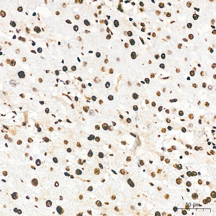
Immunohistochemistry analysis of ILF3 in paraffin-embedded mouse brain tissue using ILF3 Rabbit monoclonal antibody (STJ11102341) at a dilution of 1:200 (40x lens). High pressure antigen retrieval was performed with 0. 01 M citrate buffer (pH 6. 0) prior to immunohistochemistry staining.
-
Immunohistochemistry of paraffin-embedded rat testis using ILF3 rabbit monoclonal antibody (STJ11102341) at dilution of 1:100 (40x lens).
-
Immunohistochemistry analysis of ILF3 in paraffin-embedded human liver tissue using ILF3 Rabbit monoclonal antibody (STJ11102341) at a dilution of 1:200 (40x lens). High pressure antigen retrieval was performed with 0. 01 M citrate buffer (pH 6. 0) prior to immunohistochemistry staining.
-
Immunohistochemistry of paraffin-embedded human placenta using ILF3 rabbit monoclonal antibody (STJ11102341) at dilution of 1:100 (40x lens).
-
Immunohistochemistry analysis of ILF3 in paraffin-embedded rat liver tissue using ILF3 Rabbit monoclonal antibody (STJ11102341) at a dilution of 1:200 (40x lens). High pressure antigen retrieval was performed with 0. 01 M citrate buffer (pH 6. 0) prior to immunohistochemistry staining.
-
Immunohistochemistry of paraffin-embedded mouse kidney using ILF3 rabbit monoclonal antibody (STJ11102341) at dilution of 1:100 (40x lens).
-
Immunohistochemistry analysis of ILF3 in paraffin-embedded mouse colon tissue using ILF3 Rabbit monoclonal antibody (STJ11102341) at a dilution of 1:200 (40x lens). High pressure antigen retrieval was performed with 0. 01 M citrate buffer (pH 6. 0) prior to immunohistochemistry staining.
-
Immunohistochemistry analysis of ILF3 in paraffin-embedded rat colon tissue using ILF3 Rabbit monoclonal antibody (STJ11102341) at a dilution of 1:200 (40x lens). High pressure antigen retrieval was performed with 0. 01 M citrate buffer (pH 6. 0) prior to immunohistochemistry staining.
-
Immunohistochemistry analysis of ILF3 in paraffin-embedded mouse testis tissue using ILF3 Rabbit monoclonal antibody (STJ11102341) at a dilution of 1:200 (40x lens). High pressure antigen retrieval was performed with 0. 01 M citrate buffer (pH 6. 0) prior to immunohistochemistry staining.
-
Immunohistochemistry analysis of ILF3 in paraffin-embedded mouse lung tissue using ILF3 Rabbit monoclonal antibody (STJ11102341) at a dilution of 1:200 (40x lens). High pressure antigen retrieval was performed with 0. 01 M citrate buffer (pH 6. 0) prior to immunohistochemistry staining.
-
Immunohistochemistry analysis of ILF3 in paraffin-embedded human thyroid cancer tissue using ILF3 Rabbit monoclonal antibody (STJ11102341) at a dilution of 1:200 (40x lens). High pressure antigen retrieval was performed with 0. 01 M citrate buffer (pH 6. 0) prior to immunohistochemistry staining.
-
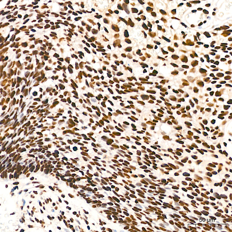
Immunohistochemistry analysis of ILF3 in paraffin-embedded human cervix cancer tissue using ILF3 Rabbit monoclonal antibody (STJ11102341) at a dilution of 1:200 (40x lens). High pressure antigen retrieval was performed with 0. 01 M citrate buffer (pH 6. 0) prior to immunohistochemistry staining.
-
Immunohistochemistry analysis of ILF3 in paraffin-embedded human colon tissue using ILF3 Rabbit monoclonal antibody (STJ11102341) at a dilution of 1:200 (40x lens). High pressure antigen retrieval was performed with 0. 01 M citrate buffer (pH 6. 0) prior to immunohistochemistry staining.
-
Immunohistochemistry analysis of ILF3 in paraffin-embedded human colon carcinoma tissue using ILF3 Rabbit monoclonal antibody (STJ11102341) at a dilution of 1:200 (40x lens). High pressure antigen retrieval was performed with 0. 01 M citrate buffer (pH 6. 0) prior to immunohistochemistry staining.
-
Western blot analysis of various lysates using ILF3 Rabbit monoclonal antibody (STJ11102341) at 1:3000 dilution. Secondary antibody: HRP Goat Anti-Rabbit IgG (H+L) (STJS000856) at 1:10000 dilution. Lysates/proteins: 25 Mu g per lane. Blocking buffer: 3% nonfat dry milk in TBST. Detection: ECL Basic Kit. Exposure time: 1s.


