| Host: |
Rabbit |
| Applications: |
WB/IP |
| Reactivity: |
Human/Mouse/Rat |
| Note: |
STRICTLY FOR FURTHER SCIENTIFIC RESEARCH USE ONLY (RUO). MUST NOT TO BE USED IN DIAGNOSTIC OR THERAPEUTIC APPLICATIONS. |
| Short Description: |
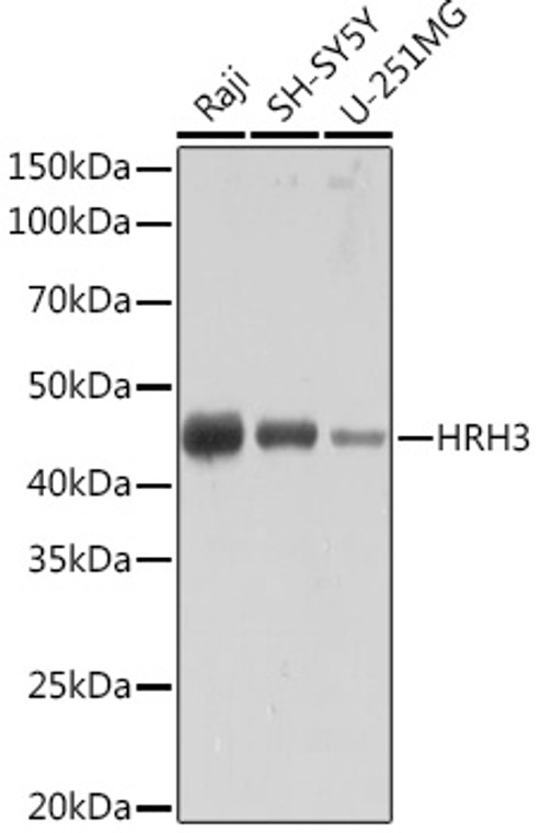
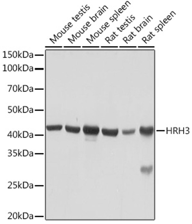
Rabbit monoclonal antibody anti-HRH3 (1-100) is suitable for use in Western Blot and Immunoprecipitation research applications. |
| Clonality: |
Monoclonal |
| Clone ID: |
S6MR |
| Conjugation: |
Unconjugated |
| Isotype: |
IgG |
| Formulation: |
PBS with 0.02% Sodium Azide, 0.05% BSA, 50% Glycerol, pH7.3. |
| Purification: |
Affinity purification |
| Dilution Range: |
WB 1:500-1:2000IP 1:500-1:1000 |
| Storage Instruction: |
Store at-20°C for up to 1 year from the date of receipt, and avoid repeat freeze-thaw cycles. |
| Gene Symbol: |
HRH3 |
| Gene ID: |
11255 |
| Uniprot ID: |
HRH3_HUMAN |
| Immunogen Region: |
1-100 |
| Immunogen: |
A synthetic peptide corresponding to a sequence within amino acids 1-100 of human HRH3 (Q9Y5N1). |
| Immunogen Sequence: |
MERAPPDGPLNASGALAGEA AAAGGARGFSAAWTAVLAAL MALLIVATVLGNALVMLAFV ADSSLRTQNNFFLLNLAISD FLVGAFCIPLYVPYVLTGRW |
| Tissue Specificity | Expressed predominantly in the CNS, with the greatest expression in the thalamus and caudate nucleus. The various isoforms are mainly coexpressed in brain, but their relative expression level varies in a region-specific manner. Isoform 3 and isoform 7 are highly expressed in the thalamus, caudate nucleus and cerebellum while isoform 5 and isoform 6 show a poor expression. Isoform 5 and isoform 6 show a high expression in the amygdala, substantia nigra, cerebral cortex and hypothalamus. Isoform 7 is not found in hypothalamus or substantia nigra. |
| Function | The H3 subclass of histamine receptors could mediate the histamine signals in CNS and peripheral nervous system. Signals through the inhibition of adenylate cyclase and displays high constitutive activity (spontaneous activity in the absence of agonist). Agonist stimulation of isoform 3 neither modified adenylate cyclase activity nor induced intracellular calcium mobilization. |
| Protein Name | Histamine H3 ReceptorH3rHh3rG-Protein Coupled Receptor 97 |
| Database Links | Reactome: R-HSA-390650 |
| Cellular Localisation | Cell MembraneMulti-Pass Membrane Protein |
| Alternative Antibody Names | Anti-Histamine H3 Receptor antibodyAnti-H3r antibodyAnti-Hh3r antibodyAnti-G-Protein Coupled Receptor 97 antibodyAnti-HRH3 antibodyAnti-GPCR97 antibody |
Information sourced from Uniprot.org
12 months for antibodies. 6 months for ELISA Kits. Please see website T&Cs for further guidance


