| Host: |
Rabbit |
| Applications: |
WB |
| Reactivity: |
Human/Mouse |
| Note: |
STRICTLY FOR FURTHER SCIENTIFIC RESEARCH USE ONLY (RUO). MUST NOT TO BE USED IN DIAGNOSTIC OR THERAPEUTIC APPLICATIONS. |
| Short Description: |
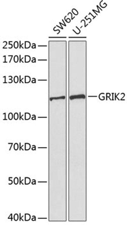
Rabbit polyclonal antibody anti-GRIK2 (30-300) is suitable for use in Western Blot research applications. |
| Clonality: |
Polyclonal |
| Conjugation: |
Unconjugated |
| Isotype: |
IgG |
| Formulation: |
PBS with 0.02% Sodium Azide, 50% Glycerol, pH7.3. |
| Purification: |
Affinity purification |
| Dilution Range: |
WB 1:500-1:2000 |
| Storage Instruction: |
Store at-20°C for up to 1 year from the date of receipt, and avoid repeat freeze-thaw cycles. |
| Gene Symbol: |
GRIK2 |
| Gene ID: |
2898 |
| Uniprot ID: |
GRIK2_HUMAN |
| Immunogen Region: |
30-300 |
| Immunogen: |
Recombinant fusion protein containing a sequence corresponding to amino acids 30-300 of human GRIK2 (NP_068775.1). |
| Immunogen Sequence: |
QGTTHVLRFGGIFEYVESGP MGAEELAFRFAVNTINRNRT LLPNTTLTYDTQKINLYDSF EASKKACDQLSLGVAAIFGP SHSSSANAVQSICNALGVPH IQTRWKHQVSDNKDSFYVSL YPDFSSLSRAILDLVQFFKW KTVTVVYDDSTGLIRLQELI KAPSRYNLRLKIRQLPADTK DAKPLLKEMKRGKEFHVIFD CSHEMAAGILKQALAMGMMT EYYHYIFTTLDLFALDVEP |
| Tissue Specificity | Expression is higher in cerebellum than in cerebral cortex. |
| Post Translational Modifications | Sumoylation mediates kainate receptor-mediated endocytosis and regulates synaptic transmission. Sumoylation is enhanced by PIAS3 and desumoylated by SENP1. Ubiquitinated. Ubiquitination regulates the GRIK2 levels at the synapse by leading kainate receptor degradation through proteasome. Phosphorylated by PKC at Ser-868 upon agonist activation, this directly enhance sumoylation. |
| Function | Ionotropic glutamate receptor. L-glutamate acts as an excitatory neurotransmitter at many synapses in the central nervous system. Binding of the excitatory neurotransmitter L-glutamate induces a conformation change, leading to the opening of the cation channel, and thereby converts the chemical signal to an electrical impulse. The receptor then desensitizes rapidly and enters a transient inactive state, characterized by the presence of bound agonist. Modulates cell surface expression of NETO2. Independent of its ionotropic glutamate receptor activity, acts as a thermoreceptor conferring sensitivity to cold temperatures. Functions in dorsal root ganglion neurons. |
| Protein Name | Glutamate Receptor Ionotropic - Kainate 2Gluk2Excitatory Amino Acid Receptor 4Eaa4Glutamate Receptor 6Glur-6Glur6 |
| Database Links | Reactome: R-HSA-451307Reactome: R-HSA-451308 |
| Cellular Localisation | Cell MembraneMulti-Pass Membrane ProteinPostsynaptic Cell Membrane |
| Alternative Antibody Names | Anti-Glutamate Receptor Ionotropic - Kainate 2 antibodyAnti-Gluk2 antibodyAnti-Excitatory Amino Acid Receptor 4 antibodyAnti-Eaa4 antibodyAnti-Glutamate Receptor 6 antibodyAnti-Glur-6 antibodyAnti-Glur6 antibodyAnti-GRIK2 antibodyAnti-GLUR6 antibody |
Information sourced from Uniprot.org
12 months for antibodies. 6 months for ELISA Kits. Please see website T&Cs for further guidance


