| Host: |
Rabbit |
| Applications: |
WB/IHC/IF |
| Reactivity: |
Human/Mouse/Rat |
| Note: |
STRICTLY FOR FURTHER SCIENTIFIC RESEARCH USE ONLY (RUO). MUST NOT TO BE USED IN DIAGNOSTIC OR THERAPEUTIC APPLICATIONS. |
| Short Description: |
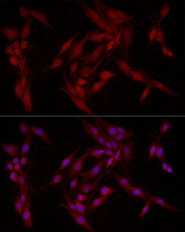
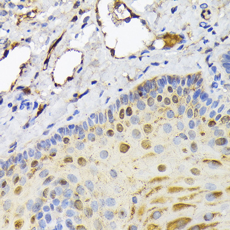
Rabbit polyclonal antibody anti-FABP5 (1-135) is suitable for use in Western Blot, Immunohistochemistry and Immunofluorescence research applications. |
| Clonality: |
Polyclonal |
| Conjugation: |
Unconjugated |
| Isotype: |
IgG |
| Formulation: |
PBS with 0.05% Proclin300, 50% Glycerol, pH7.3. |
| Purification: |
Affinity purification |
| Dilution Range: |
WB 1:500-1:1000IHC-P 1:50-1:200IF/ICC 1:50-1:200 |
| Storage Instruction: |
Store at-20°C for up to 1 year from the date of receipt, and avoid repeat freeze-thaw cycles. |
| Gene Symbol: |
FABP5 |
| Gene ID: |
2171 |
| Uniprot ID: |
FABP5_HUMAN |
| Immunogen Region: |
1-135 |
| Immunogen: |
Recombinant fusion protein containing a sequence corresponding to amino acids 1-135 of human FABP5 (NP_001435.1). |
| Immunogen Sequence: |
MATVQQLEGRWRLVDSKGFD EYMKELGVGIALRKMGAMAK PDCIITCDGKNLTIKTESTL KTTQFSCTLGEKFEETTADG RKTQTVCNFTDGALVQHQEW DGKESTITRKLKDGKLVVEC VMNNVTCTRIYEKVE |
| Tissue Specificity | Keratinocytes.highly expressed in psoriatic skin. Expressed in brain gray matter. |
| Function | Intracellular carrier for long-chain fatty acids and related active lipids, such as endocannabinoids, that regulate the metabolism and actions of the ligands they bind. In addition to the cytosolic transport, selectively delivers specific fatty acids from the cytosol to the nucleus, wherein they activate nuclear receptors. Delivers retinoic acid to the nuclear receptor peroxisome proliferator-activated receptor delta.which promotes proliferation and survival. May also serve as a synaptic carrier of endocannabinoid at central synapses and thus controls retrograde endocannabinoid signaling. Modulates inflammation by regulating PTGES induction via NF-kappa-B activation, and prostaglandin E2 (PGE2) biosynthesis during inflammation. May be involved in keratinocyte differentiation. |
| Protein Name | Fatty Acid-Binding Protein 5Epidermal-Type Fatty Acid-Binding ProteinE-FabpFatty Acid-Binding Protein - EpidermalPsoriasis-Associated Fatty Acid-Binding Protein HomologPa-Fabp |
| Database Links | Reactome: R-HSA-163560Reactome: R-HSA-5362517Reactome: R-HSA-6798695 |
| Cellular Localisation | CytoplasmNucleusSynapsePostsynaptic DensitySecretedLocalizes Primarily To The CytoplasmUpon Certain Ligand BindingA Conformation Change Exposes A Nuclear Localization Motif And The Protein Is Transported Into NucleusSecreted By AstrocytesBut Not By Neurons |
| Alternative Antibody Names | Anti-Fatty Acid-Binding Protein 5 antibodyAnti-Epidermal-Type Fatty Acid-Binding Protein antibodyAnti-E-Fabp antibodyAnti-Fatty Acid-Binding Protein - Epidermal antibodyAnti-Psoriasis-Associated Fatty Acid-Binding Protein Homolog antibodyAnti-Pa-Fabp antibodyAnti-FABP5 antibody |
Information sourced from Uniprot.org
12 months for antibodies. 6 months for ELISA Kits. Please see website T&Cs for further guidance


