| Host: |
Rabbit |
| Applications: |
WB |
| Reactivity: |
Human/Mouse |
| Note: |
STRICTLY FOR FURTHER SCIENTIFIC RESEARCH USE ONLY (RUO). MUST NOT TO BE USED IN DIAGNOSTIC OR THERAPEUTIC APPLICATIONS. |
| Short Description: |
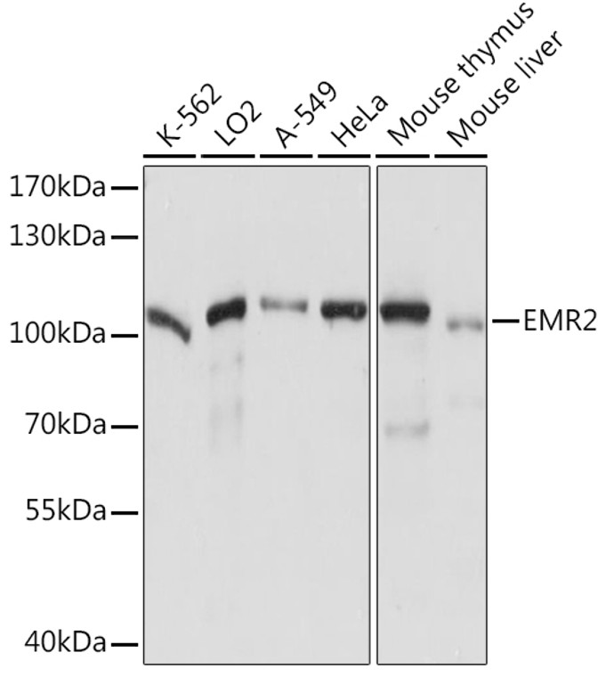
Rabbit polyclonal antibody anti-EMR2 (300-500) is suitable for use in Western Blot research applications. |
| Clonality: |
Polyclonal |
| Conjugation: |
Unconjugated |
| Isotype: |
IgG |
| Formulation: |
PBS with 0.01% Thimerosal, 50% Glycerol, pH7.3. |
| Purification: |
Affinity purification |
| Dilution Range: |
WB 1:500-1:2000 |
| Storage Instruction: |
Store at-20°C for up to 1 year from the date of receipt, and avoid repeat freeze-thaw cycles. |
| Gene Symbol: |
ADGRE2 |
| Gene ID: |
30817 |
| Uniprot ID: |
AGRE2_HUMAN |
| Immunogen Region: |
300-500 |
| Immunogen: |
Recombinant fusion protein containing a sequence corresponding to amino acids 300-500 of human EMR2 (NP_038475.2). |
| Immunogen Sequence: |
TIQSILQALDELLEAPGDLE TLPRLQQHCVASHLLDGLED VLRGLSKNLSNGLLNFSYPA GTELSLEVQKQVDRSVTLRQ NQAVMQLDWNQAQKSGDPGP SVVGLVSIPGMGKLLAEAPL VLEPEKQMLLHETHQGLLQD GSPILLSDVISAFLSNNDTQ NLSSPVTFTFSHRSVIPRQK VLCVFWEHGQNGCGHWATTG C |
| Tissue Specificity | Expression is restricted to myeloid cells. Highest expression was found in peripheral blood leukocytes, followed by spleen and lymph nodes, with intermediate to low levels in thymus, bone marrow, fetal liver, placenta, and lung, and no expression in heart, brain, skeletal muscle, kidney, or pancreas. Expression is also detected in monocyte/macrophage and Jurkat cell lines but not in other cell lines tested. High expression in mast cells. |
| Post Translational Modifications | Autoproteolytically cleaved into 2 subunits, an extracellular alpha subunit and a seven-transmembrane beta subunit. |
| Function | Cell surface receptor that binds to the chondroitin sulfate moiety of glycosaminoglycan chains and promotes cell attachment. Promotes granulocyte chemotaxis, degranulation and adhesion. In macrophages, promotes the release of inflammatory cytokines, including IL8 and TNF. Signals probably through G-proteins. Is a regulator of mast cell degranulation. |
| Protein Name | Adhesion G Protein-Coupled Receptor E2Egf-Like Module Receptor 2Egf-Like Module-Containing Mucin-Like Hormone Receptor-Like 2Cd Antigen Cd312 |
| Database Links | Reactome: R-HSA-373080 |
| Cellular Localisation | Cell MembraneMulti-Pass Membrane ProteinCell ProjectionRuffle MembraneLocalized At The Leading Edge Of Migrating Cells |
| Alternative Antibody Names | Anti-Adhesion G Protein-Coupled Receptor E2 antibodyAnti-Egf-Like Module Receptor 2 antibodyAnti-Egf-Like Module-Containing Mucin-Like Hormone Receptor-Like 2 antibodyAnti-Cd Antigen Cd312 antibodyAnti-ADGRE2 antibodyAnti-EMR2 antibody |
Information sourced from Uniprot.org
12 months for antibodies. 6 months for ELISA Kits. Please see website T&Cs for further guidance


