| Host: |
Goat |
| Applications: |
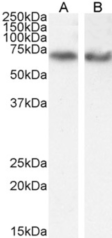
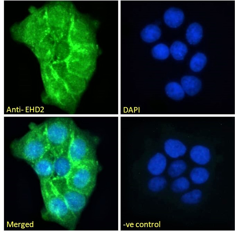
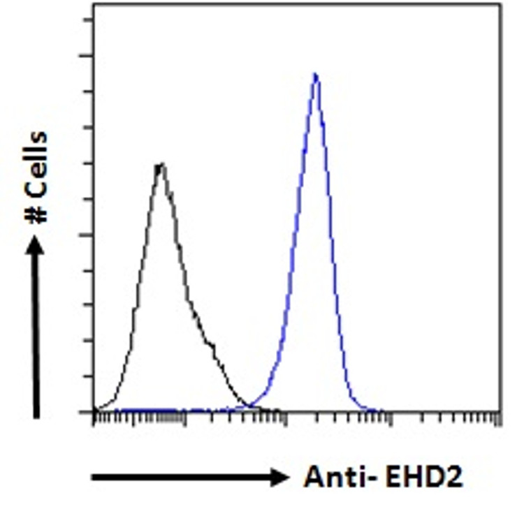
Pep-ELISA/WB/IF/FC |
| Reactivity: |
Human/Mouse/Rat/Dog/Cow |
| Note: |
STRICTLY FOR FURTHER SCIENTIFIC RESEARCH USE ONLY (RUO). MUST NOT TO BE USED IN DIAGNOSTIC OR THERAPEUTIC APPLICATIONS. |
| Short Description: |
Goat polyclonal antibody anti-EHD2 (C-Term) is suitable for use in ELISA, Western Blot, Immunofluorescence and Flow Cytometry research applications. |
| Clonality: |
Polyclonal |
| Conjugation: |
Unconjugated |
| Isotype: |
IgG |
| Formulation: |
0.5 mg/ml in Tris saline, 0.02% sodium azide, pH7.3 with 0.5% bovine serum albumin. NA |
| Purification: |
Purified from goat serum by ammonium sulphate precipitation followed by antigen affinity chromatography using the immunizing peptide. |
| Concentration: |
0.5 mg/mL |
| Dilution Range: |
WB-0.1-0.3µg/mlIF-Strong expression of the protein seen in the plasma membrane of A431 cells and the nuclei of HeLa cells. 10µg/mlELISA-antibody detection limit dilution 1:64000. |
| Storage Instruction: |
Store at-20°C on receipt and minimise freeze-thaw cycles. |
| Gene Symbol: |
EHD2 |
| Gene ID: |
30846 |
| Uniprot ID: |
EHD2_HUMAN |
| Immunogen Region: |
C-Term |
| Accession Number: |
NP_055416.2 |
| Specificity: |
This antibody is expected to recognise EHD1 protein as well as EHD2. |
| Immunogen Sequence: |
CRLVPPSKRRHKGSA |
| Function | ATP- and membrane-binding protein that controls membrane reorganization/tubulation upon ATP hydrolysis. Plays a role in membrane trafficking between the plasma membrane and endosomes. Important for the internalization of GLUT4. Required for fusion of myoblasts to skeletal muscle myotubes. Required for normal translocation of FER1L5 to the plasma membrane. Regulates the equilibrium between cell surface-associated and cell surface-dissociated caveolae by constraining caveolae at the cell membrane. |
| Protein Name | Eh Domain-Containing Protein 2Past Homolog 2 |
| Database Links | Reactome: R-HSA-983231 |
| Cellular Localisation | Cell MembranePeripheral Membrane ProteinCytoplasmic SideMembraneCaveolaEndosome MembraneCytoplasmCytosolColocalizes With Glut4 In Intracellular Tubulovesicular Structures That Are Associated With Cortical F-ActinColocalizes With Fer1l5 At Plasma Membrane In Myoblasts And Myotubes |
| Alternative Antibody Names | Anti-Eh Domain-Containing Protein 2 antibodyAnti-Past Homolog 2 antibodyAnti-EHD2 antibodyAnti-PAST2 antibody |
Information sourced from Uniprot.org
12 months for antibodies. 6 months for ELISA Kits. Please see website T&Cs for further guidance


