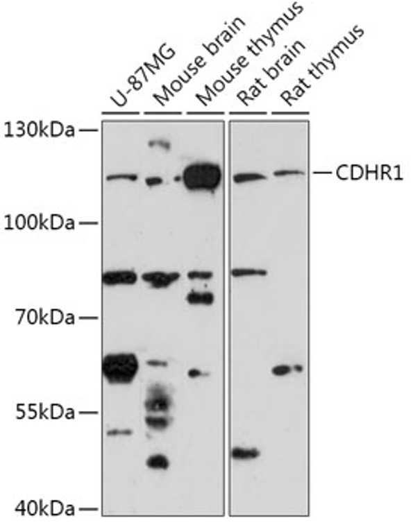
| Host: |
Rabbit |
| Applications: |
WB |
| Reactivity: |
Human/Mouse/Rat |
| Note: |
STRICTLY FOR FURTHER SCIENTIFIC RESEARCH USE ONLY (RUO). MUST NOT TO BE USED IN DIAGNOSTIC OR THERAPEUTIC APPLICATIONS. |
| Short Description: |
Rabbit polyclonal antibody anti-CDHR1 (20-300) is suitable for use in Western Blot research applications. |
| Clonality: |
Polyclonal |
| Conjugation: |
Unconjugated |
| Isotype: |
IgG |
| Formulation: |
PBS with 0.01% Thimerosal, 50% Glycerol, pH7.3. |
| Purification: |
Affinity purification |
| Dilution Range: |
WB 1:500-1:2000 |
| Storage Instruction: |
Store at-20°C for up to 1 year from the date of receipt, and avoid repeat freeze-thaw cycles. |
| Gene Symbol: |
CDHR1 |
| Gene ID: |
92211 |
| Uniprot ID: |
CDHR1_HUMAN |
| Immunogen Region: |
20-300 |
| Immunogen: |
Recombinant fusion protein containing a sequence corresponding to amino acids 20-300 of human CDHR1 (NP_001165442.1). |
| Immunogen Sequence: |
QANFAPHFFDNGVGSTNGNM ALFSLPEDTPVGSHVYTLNG TDPEGDPISYHISFDPSTRS VFSVDPTFGNITLVEELDRE REDEIEAIISISDGLNLVAE KVVILVTDANDEAPRFIQEP YVALVPEDIPAGSIIFKVHA VDRDTGSGGSVTYFLQNLHS PFAVDRHSGVLRLQAGATLD YERSRTHYITVVAKDGGGRL HGADVVFSATTTVTVNVEDV QDMAPVFVGTPYYGYVYED |
| Post Translational Modifications | Undergoes proteolytic cleavage.produces a soluble 95 kDa N-terminal fragment and a 25 kDa cell-associated C-terminal fragment. |
| Function | Potential calcium-dependent cell-adhesion protein. May be required for the structural integrity of the outer segment (OS) of photoreceptor cells. |
| Protein Name | Cadherin-Related Family Member 1Photoreceptor CadherinPrcadProtocadherin-21 |
| Cellular Localisation | Cell MembraneSingle-Pass Membrane ProteinLocalized At The Junction Between The Inner And Outer Segments Of Rod And Cone Photoreceptors CellsConfined To The Base Of The OsLocalized On The Edges Of Nascent Evaginating Disks On The Side Of The Os Opposite The Connecting CiliumExpressed At Postnatal Day 2 At The Apical Tip Of The Rod Photoreceptor CellsThe Site Of The Developing OsColocalized With Rhodopsin Between Postnatal Days 2 And 9 At The Base Of The Growing Os Region |
| Alternative Antibody Names | Anti-Cadherin-Related Family Member 1 antibodyAnti-Photoreceptor Cadherin antibodyAnti-Prcad antibodyAnti-Protocadherin-21 antibodyAnti-CDHR1 antibodyAnti-KIAA1775 antibodyAnti-PCDH21 antibodyAnti-PRCAD antibody |
Information sourced from Uniprot.org
12 months for antibodies. 6 months for ELISA Kits. Please see website T&Cs for further guidance


