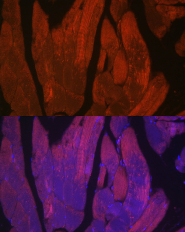
| Host: |
Rabbit |
| Applications: |
IF/ICC/ELISA |
| Reactivity: |
Mouse/Rat |
| Note: |
STRICTLY FOR FURTHER SCIENTIFIC RESEARCH USE ONLY (RUO). MUST NOT TO BE USED IN DIAGNOSTIC OR THERAPEUTIC APPLICATIONS. |
| Clonality: |
Polyclonal |
| Conjugation: |
Unconjugated |
| Isotype: |
IgG |
| Formulation: |
PBS with 0.05% Proclin300, 50% Glycerol, pH 7.3. |
| Purification: |
Affinity purification |
| Concentration: |
Lot specific |
| Dilution Range: |
IF/ICC:1:50-1:200ELISA:Recommended starting concentration is 1 Mu g/mL. Please optimize the concentration based on your specific assay requirements. |
| Storage Instruction: |
Store at-20°C for up to 1 year from the date of receipt, and avoid repeat freeze-thaw cycles. |
| Gene Symbol: |
CACNA1S |
| Gene ID: |
779 |
| Uniprot ID: |
CAC1S_HUMAN |
| Immunogen Region: |
900-1000 aa |
| Specificity: |
Recombinant fusion protein containing a sequence corresponding to amino acids 900-1000 of human CACNA1S. (NP_000060.2). |
| Immunogen Sequence: |
RVLRPLRAINRAKGLKHVVQ CMFVAISTIGNIVLVTTLLQ FMFACIGVQLFKGKFFRCTD LSKMTEEECRGYYYVYKDGD PMQIELRHREWVHSDFHFDN V |
| Tissue Specificity | Skeletal muscle specific. |
| Post Translational Modifications | The alpha-1S subunit is found in two isoforms in the skeletal muscle: a minor form of 212 kDa containing the complete amino acid sequence, and a major form of 190 kDa derived from the full-length form by post-translational proteolysis close to Phe-1690. Phosphorylated. Phosphorylation by PKA activates the calcium channel. Both the minor and major forms are phosphorylated in vitro by PKA. Phosphorylation at Ser-1575 is involved in beta-adrenergic-mediated regulation of the channel. |
| Function | Pore-forming, alpha-1S subunit of the voltage-gated calcium channel that gives rise to L-type calcium currents in skeletal muscle. Calcium channels containing the alpha-1S subunit play an important role in excitation-contraction coupling in skeletal muscle via their interaction with RYR1, which triggers Ca(2+) release from the sarcoplasmic reticulum and ultimately results in muscle contraction. Long-lasting (L-type) calcium channels belong to the 'high-voltage activated' (HVA) group. |
| Protein Name | Voltage-Dependent L-Type Calcium Channel Subunit Alpha-1sCalcium Channel - L Type - Alpha-1 Polypeptide - Isoform 3 - Skeletal MuscleVoltage-Gated Calcium Channel Subunit Alpha Cav1.1 |
| Database Links | Reactome: R-HSA-419037 |
| Cellular Localisation | Cell MembraneSarcolemmaT-TubuleMulti-Pass Membrane Protein |
| Alternative Antibody Names | Anti-Voltage-Dependent L-Type Calcium Channel Subunit Alpha-1s antibodyAnti-Calcium Channel - L Type - Alpha-1 Polypeptide - Isoform 3 - Skeletal Muscle antibodyAnti-Voltage-Gated Calcium Channel Subunit Alpha Cav1.1 antibodyAnti-CACNA1S antibodyAnti-CACH1 antibodyAnti-CACN1 antibodyAnti-CACNL1A3 antibody |
Information sourced from Uniprot.org
12 months for antibodies. 6 months for ELISA Kits. Please see website T&Cs for further guidance


