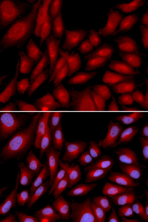
| Host: |
Rabbit |
| Applications: |
WB/IF |
| Reactivity: |
Human/Mouse |
| Note: |
STRICTLY FOR FURTHER SCIENTIFIC RESEARCH USE ONLY (RUO). MUST NOT TO BE USED IN DIAGNOSTIC OR THERAPEUTIC APPLICATIONS. |
| Short Description: |
Rabbit polyclonal antibody anti-BAG1 (1-230) is suitable for use in Western Blot and Immunofluorescence research applications. |
| Clonality: |
Polyclonal |
| Conjugation: |
Unconjugated |
| Isotype: |
IgG |
| Formulation: |
PBS with 0.02% Sodium Azide, 50% Glycerol, pH7.3. |
| Purification: |
Affinity purification |
| Dilution Range: |
WB 1:500-1:2000IF/ICC 1:20-1:100 |
| Storage Instruction: |
Store at-20°C for up to 1 year from the date of receipt, and avoid repeat freeze-thaw cycles. |
| Gene Symbol: |
BAG1 |
| Gene ID: |
573 |
| Uniprot ID: |
BAG1_HUMAN |
| Immunogen Region: |
1-230 |
| Immunogen: |
Recombinant fusion protein containing a sequence corresponding to amino acids 1-230 of human BAG1 (NP_001165886.1). |
| Immunogen Sequence: |
MNRSQEVTRDEESTRSEEVT REEMAAAGLTVTVTHSNEKH DLHVTSQQGSSEPVVQDLAQ VVEEVIGVPQSFQKLIFKGK SLKEMETPLSALGIQDGCRV MLIGKKNSPQEEVELKKLKH LEKSVEKIADQLEELNKELT GIQQGFLPKDLQAEALCKLD RRVKATIEQFMKILEEIDTL ILPENFKDSRLKRKGLVKKV QAFLAECDTVEQNICQETER LQSTNFALAE |
| Tissue Specificity | Isoform 4 is the most abundantly expressed isoform. It is ubiquitously expressed throughout most tissues, except the liver, colon, breast and uterine myometrium. Isoform 1 is expressed in the ovary and testis. Isoform 4 is expressed in several types of tumor cell lines, and at consistently high levels in leukemia and lymphoma cell lines. Isoform 1 is expressed in the prostate, breast and leukemia cell lines. Isoform 3 is the least abundant isoform in tumor cell lines (at protein level). |
| Post Translational Modifications | Ubiquitinated.mediated by SIAH1 or SIAH2 and leading to its subsequent proteasomal degradation. |
| Function | Co-chaperone for HSP70 and HSC70 chaperone proteins. Acts as a nucleotide-exchange factor (NEF) promoting the release of ADP from the HSP70 and HSC70 proteins thereby triggering client/substrate protein release. Nucleotide release is mediated via its binding to the nucleotide-binding domain (NBD) of HSPA8/HSC70 where as the substrate release is mediated via its binding to the substrate-binding domain (SBD) of HSPA8/HSC70. Inhibits the pro-apoptotic function of PPP1R15A, and has anti-apoptotic activity. Markedly increases the anti-cell death function of BCL2 induced by various stimuli. |
| Protein Name | Bag Family Molecular Chaperone Regulator 1Bag-1Bcl-2-Associated Athanogene 1 |
| Database Links | Reactome: R-HSA-3371453 |
| Cellular Localisation | Isoform 1: NucleusCytoplasmIsoform 1 Localizes Predominantly To The NucleusIsoform 2: CytoplasmNucleusIsoform 2 Localizes To The Cytoplasm And Shuttles Into The Nucleus In Response To Heat ShockIsoform 4: CytoplasmIsoform 4 Localizes Predominantly To The CytoplasmThe Cellular Background In Which It Is Expressed Can Influence Whether It Resides Primarily In The Cytoplasm Or Is Also Found In The NucleusIn The Presence Of Bcl2Localizes To Intracellular Membranes (What Appears To Be The Nuclear Envelope And Perinuclear Membranes) As Well As Punctate Cytosolic Structures Suggestive Of Mitochondria |
| Alternative Antibody Names | Anti-Bag Family Molecular Chaperone Regulator 1 antibodyAnti-Bag-1 antibodyAnti-Bcl-2-Associated Athanogene 1 antibodyAnti-BAG1 antibodyAnti-HAP antibody |
Information sourced from Uniprot.org
12 months for antibodies. 6 months for ELISA Kits. Please see website T&Cs for further guidance


