| Host: |
Rabbit |
| Applications: |
WB/IHC |
| Reactivity: |
Human/Mouse/Rat |
| Note: |
STRICTLY FOR FURTHER SCIENTIFIC RESEARCH USE ONLY (RUO). MUST NOT TO BE USED IN DIAGNOSTIC OR THERAPEUTIC APPLICATIONS. |
| Short Description: |
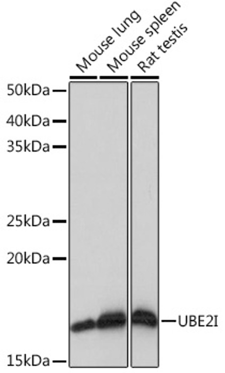
Rabbit monoclonal antibody anti-UBE2I (1-100) is suitable for use in Western Blot and Immunohistochemistry research applications. |
| Clonality: |
Monoclonal |
| Clone ID: |
S9MR |
| Conjugation: |
Unconjugated |
| Isotype: |
IgG |
| Formulation: |
PBS with 0.02% Sodium Azide, 0.05% BSA, 50% Glycerol, pH7.3. |
| Purification: |
Affinity purification |
| Dilution Range: |
WB 1:500-1:1000IHC-P 1:50-1:200 |
| Storage Instruction: |
Store at-20°C for up to 1 year from the date of receipt, and avoid repeat freeze-thaw cycles. |
| Gene Symbol: |
UBE2I |
| Gene ID: |
7329 |
| Uniprot ID: |
UBC9_HUMAN |
| Immunogen Region: |
1-100 |
| Immunogen: |
A synthetic peptide corresponding to a sequence within amino acids 1-100 of human UBE2I (P63279). |
| Immunogen Sequence: |
MSGIALSRLAQERKAWRKDH PFGFVAVPTKNPDGTMNLMN WECAIPGKKGTPWEGGLFKL RMLFKDDYPSSPPKCKFEPP LFHPNVYPSGTVCLSILEED |
| Tissue Specificity | Expressed in heart, skeletal muscle, pancreas, kidney, liver, lung, placenta and brain. Also expressed in testis and thymus. |
| Post Translational Modifications | Phosphorylation at Ser-71 significantly enhances SUMOylation activity. |
| Function | Accepts the ubiquitin-like proteins SUMO1, SUMO2, SUMO3, SUMO4 and SUMO1P1/SUMO5 from the UBLE1A-UBLE1B E1 complex and catalyzes their covalent attachment to other proteins with the help of an E3 ligase such as RANBP2, CBX4 and ZNF451. Can catalyze the formation of poly-SUMO chains. Necessary for sumoylation of FOXL2 and KAT5. Essential for nuclear architecture and chromosome segregation. Sumoylates p53/TP53 at 'Lys-386'. Mediates sumoylation of ERCC6 which is essential for its transcription-coupled nucleotide excision repair activity. |
| Protein Name | Sumo-Conjugating Enzyme Ubc9Ring-Type E3 Sumo Transferase Ubc9Sumo-Protein LigaseUbiquitin Carrier Protein 9Ubiquitin Carrier Protein IUbiquitin-Conjugating Enzyme E2 IUbiquitin-Protein Ligase IP18 |
| Database Links | Reactome: R-HSA-1221632Reactome: R-HSA-196791Reactome: R-HSA-3065678Reactome: R-HSA-3108214Reactome: R-HSA-3232118Reactome: R-HSA-3232142Reactome: R-HSA-3899300Reactome: R-HSA-4085377Reactome: R-HSA-4090294Reactome: R-HSA-4551638Reactome: R-HSA-4570464Reactome: R-HSA-4615885Reactome: R-HSA-4655427Reactome: R-HSA-4755510Reactome: R-HSA-5693565Reactome: R-HSA-5693607Reactome: R-HSA-5696395Reactome: R-HSA-8866904Reactome: R-HSA-9615933Reactome: R-HSA-9683610Reactome: R-HSA-9694631Reactome: R-HSA-9735871Reactome: R-HSA-9793242 |
| Cellular Localisation | NucleusCytoplasmPerinuclear RegionMainly NuclearIn SpermatocytesLocalizes In Synaptonemal ComplexesRecruited By Bcl11a Into The Nuclear Body |
| Alternative Antibody Names | Anti-Sumo-Conjugating Enzyme Ubc9 antibodyAnti-Ring-Type E3 Sumo Transferase Ubc9 antibodyAnti-Sumo-Protein Ligase antibodyAnti-Ubiquitin Carrier Protein 9 antibodyAnti-Ubiquitin Carrier Protein I antibodyAnti-Ubiquitin-Conjugating Enzyme E2 I antibodyAnti-Ubiquitin-Protein Ligase I antibodyAnti-P18 antibodyAnti-UBE2I antibodyAnti-UBC9 antibodyAnti-UBCE9 antibody |
Information sourced from Uniprot.org
12 months for antibodies. 6 months for ELISA Kits. Please see website T&Cs for further guidance


