| Host: |
Rabbit |
| Applications: |
WB/IF |
| Reactivity: |
Human/Mouse/Rat |
| Note: |
STRICTLY FOR FURTHER SCIENTIFIC RESEARCH USE ONLY (RUO). MUST NOT TO BE USED IN DIAGNOSTIC OR THERAPEUTIC APPLICATIONS. |
| Short Description: |
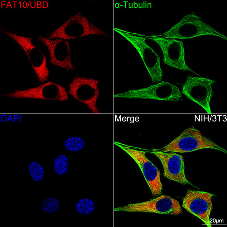
Rabbit monoclonal antibody anti-Ubiquitin D (70-150) is suitable for use in Western Blot and Immunofluorescence research applications. |
| Clonality: |
Monoclonal |
| Clone ID: |
S7MR |
| Conjugation: |
Unconjugated |
| Isotype: |
IgG |
| Formulation: |
PBS with 0.02% Sodium Azide, 0.05% BSA, 50% Glycerol, pH7.3. |
| Purification: |
Affinity purification |
| Dilution Range: |
WB 1:500-1:2000IF/ICC 1:50-1:200 |
| Storage Instruction: |
Store at-20°C for up to 1 year from the date of receipt, and avoid repeat freeze-thaw cycles. |
| Gene Symbol: |
UBD |
| Gene ID: |
10537 |
| Uniprot ID: |
UBD_HUMAN |
| Immunogen Region: |
70-150 |
| Immunogen: |
A synthetic peptide corresponding to a sequence within amino acids 70-150 of human FAT10/UBD (O15205). |
| Immunogen Sequence: |
KEKTIHLTLKVVKPSDEELP LFLVESGDEAKRHLLQVRRS SSVAQVKAMIETKTGIIPET QIVTCNGKRLEDGKMMADYG I |
| Tissue Specificity | Constitutively expressed in mature dendritic cells and B-cells. Mostly expressed in the reticuloendothelial system (e.g. thymus, spleen), the gastrointestinal system, kidney, lung and prostate gland. |
| Post Translational Modifications | Can be acetylated. |
| Function | Ubiquitin-like protein modifier which can be covalently attached to target protein and subsequently leads to their degradation by the 26S proteasome, in a NUB1-dependent manner. Probably functions as a survival factor. Conjugation ability activated by UBA6. Promotes the expression of the proteasome subunit beta type-9 (PSMB9/LMP2). Regulates TNF-alpha-induced and LPS-mediated activation of the central mediator of innate immunity NF-kappa-B by promoting TNF-alpha-mediated proteasomal degradation of ubiquitinated-I-kappa-B-alpha. Required for TNF-alpha-induced p65 nuclear translocation in renal tubular epithelial cells (RTECs). May be involved in dendritic cell (DC) maturation, the process by which immature dendritic cells differentiate into fully competent antigen-presenting cells that initiate T-cell responses. Mediates mitotic non-disjunction and chromosome instability, in long-term in vitro culture and cancers, by abbreviating mitotic phase and impairing the kinetochore localization of MAD2L1 during the prometaphase stage of the cell cycle. May be involved in the formation of aggresomes when proteasome is saturated or impaired. Mediates apoptosis in a caspase-dependent manner, especially in renal epithelium and tubular cells during renal diseases such as polycystic kidney disease and Human immunodeficiency virus (HIV)-associated nephropathy (HIVAN). |
| Protein Name | Ubiquitin DDiubiquitinUbiquitin-Like Protein Fat10 |
| Database Links | Reactome: R-HSA-8951664 |
| Cellular Localisation | NucleusCytoplasmAccumulates In Aggresomes Under Proteasome Inhibition Conditions |
| Alternative Antibody Names | Anti-Ubiquitin D antibodyAnti-Diubiquitin antibodyAnti-Ubiquitin-Like Protein Fat10 antibodyAnti-UBD antibodyAnti-FAT10 antibody |
Information sourced from Uniprot.org
12 months for antibodies. 6 months for ELISA Kits. Please see website T&Cs for further guidance


