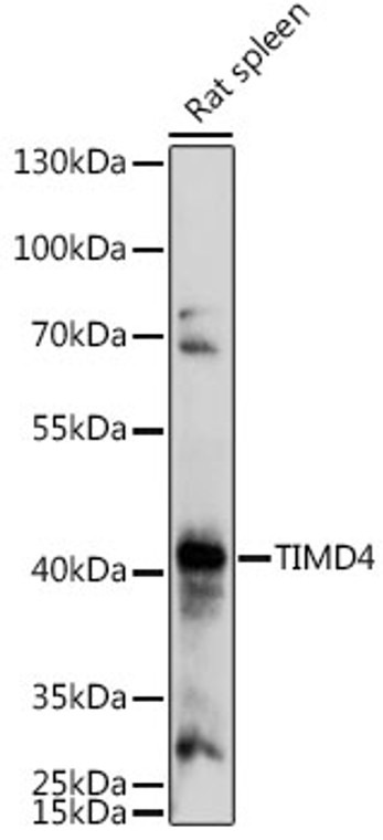
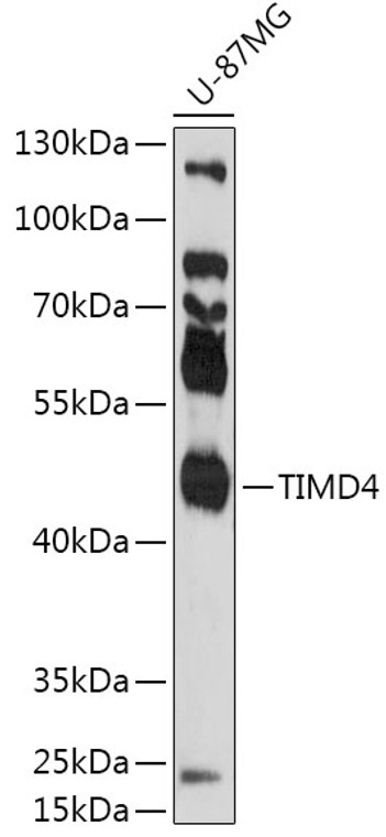
| Host: |
Rabbit |
| Applications: |
WB |
| Reactivity: |
Human/Rat |
| Note: |
STRICTLY FOR FURTHER SCIENTIFIC RESEARCH USE ONLY (RUO). MUST NOT TO BE USED IN DIAGNOSTIC OR THERAPEUTIC APPLICATIONS. |
| Short Description: |
Rabbit polyclonal antibody anti-TIMD4 (25-130) is suitable for use in Western Blot research applications. |
| Clonality: |
Polyclonal |
| Conjugation: |
Unconjugated |
| Isotype: |
IgG |
| Formulation: |
PBS with 0.01% Thimerosal, 50% Glycerol, pH7.3. |
| Purification: |
Affinity purification |
| Dilution Range: |
WB 1:500-1:1000 |
| Storage Instruction: |
Store at-20°C for up to 1 year from the date of receipt, and avoid repeat freeze-thaw cycles. |
| Gene Symbol: |
TIMD4 |
| Gene ID: |
91937 |
| Uniprot ID: |
TIMD4_HUMAN |
| Immunogen Region: |
25-130 |
| Immunogen: |
Recombinant fusion protein containing a sequence corresponding to amino acids 25-130 of human TIMD4 (NP_612388.2). |
| Immunogen Sequence: |
ETVVTEVLGHRVTLPCLYSS WSHNSNSMCWGKDQCPYSGC KEALIRTDGMRVTSRKSAKY RLQGTIPRGDVSLTILNPSE SDSGVYCCRIEVPGWFNDVK INVRLN |
| Function | Phosphatidylserine receptor that plays different role in immune response including phagocytosis of apoptotic cells and T-cell regulation. Controls T-cell activation in a bimodal fashion, decreasing the activation of naive T-cells by inducing cell cycle arrest, while increasing proliferation of activated T-cells by activating AKT1 and ERK1/2 phosphorylations and subsequent signaling pathways. Also plays a role in efferocytosis which is the process by which apoptotic cells are removed by phagocytic cells. Mechanistically, promotes the engulfment of apoptotic cells or exogenous particles by securing them to phagocytes through direct binding to phosphatidylserine present on apoptotic cells, while other engulfment receptors such as MERTK efficiently recognize apoptotic cells and mediate their ingestion. Additionally, promotes autophagy process by suppressing NLRP3 inflammasome activity via activation of LKB1/PRKAA1 pathway in a phosphatidylserine-dependent mechanism. (Microbial infection) Plays a positive role in exosome-mediated trafficking of HIV-1 virus and its entry into immune cells. |
| Protein Name | T-Cell Immunoglobulin And Mucin Domain-Containing Protein 4Timd-4T-Cell Immunoglobulin Mucin Receptor 4Tim-4T-Cell Membrane Protein 4 |
| Cellular Localisation | Cell MembraneSingle-Pass Type I Membrane ProteinSecretedExtracellular Exosome |
| Alternative Antibody Names | Anti-T-Cell Immunoglobulin And Mucin Domain-Containing Protein 4 antibodyAnti-Timd-4 antibodyAnti-T-Cell Immunoglobulin Mucin Receptor 4 antibodyAnti-Tim-4 antibodyAnti-T-Cell Membrane Protein 4 antibodyAnti-TIMD4 antibodyAnti-TIM4 antibody |
Information sourced from Uniprot.org
12 months for antibodies. 6 months for ELISA Kits. Please see website T&Cs for further guidance


