| Host: |
Rabbit |
| Applications: |
WB/IHC/IF |
| Reactivity: |
Human/Mouse/Rat |
| Note: |
STRICTLY FOR FURTHER SCIENTIFIC RESEARCH USE ONLY (RUO). MUST NOT TO BE USED IN DIAGNOSTIC OR THERAPEUTIC APPLICATIONS. |
| Short Description: |
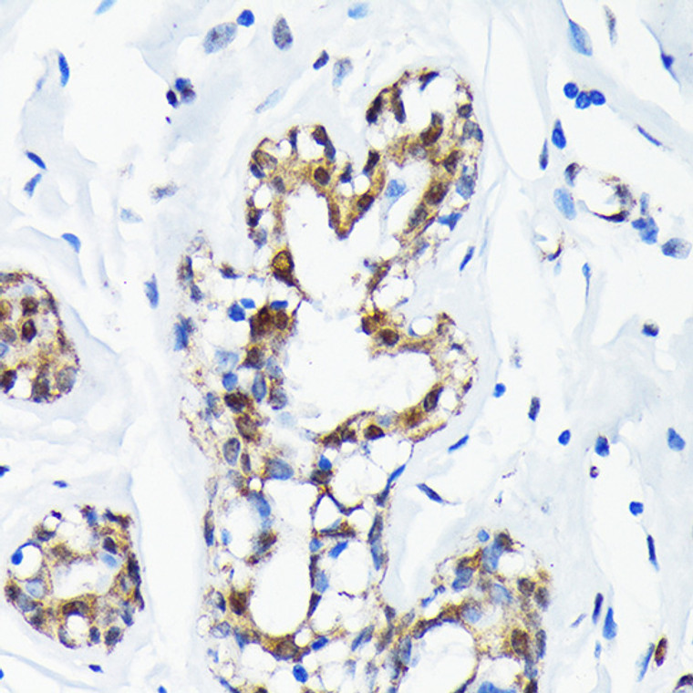
Rabbit monoclonal antibody anti-SUMO1 (1-101) is suitable for use in Western Blot, Immunohistochemistry and Immunofluorescence research applications. |
| Clonality: |
Monoclonal |
| Clone ID: |
S8MR |
| Conjugation: |
Unconjugated |
| Isotype: |
IgG |
| Formulation: |
PBS with 0.02% Sodium Azide, 0.05% BSA, 50% Glycerol, pH7.3. |
| Purification: |
Affinity purification |
| Dilution Range: |
WB 1:500-1:1000IHC-P 1:50-1:200IF/ICC 1:50-1:200 |
| Storage Instruction: |
Store at-20°C for up to 1 year from the date of receipt, and avoid repeat freeze-thaw cycles. |
| Gene Symbol: |
SUMO1 |
| Gene ID: |
7341 |
| Uniprot ID: |
SUMO1_HUMAN |
| Immunogen Region: |
1-101 |
| Immunogen: |
A synthetic peptide corresponding to a sequence within amino acids 1-101 of human Sumo 1 (P63165). |
| Immunogen Sequence: |
MSDQEAKPSTEDLGDKKEGE YIKLKVIGQDSSEIHFKVKM TTHLKKLKESYCQRQGVPMN SLRFLFEGQRIADNHTPKEL GMEEEDVIEVYQEQTGGHST V |
| Post Translational Modifications | Cleavage of precursor form by SENP1 or SENP2 is necessary for function. Polymeric SUMO1 chains undergo polyubiquitination by RNF4. |
| Function | Ubiquitin-like protein that can be covalently attached to proteins as a monomer or a lysine-linked polymer. Covalent attachment via an isopeptide bond to its substrates requires prior activation by the E1 complex SAE1-SAE2 and linkage to the E2 enzyme UBE2I, and can be promoted by E3 ligases such as PIAS1-4, RANBP2 or CBX4. This post-translational modification on lysine residues of proteins plays a crucial role in a number of cellular processes such as nuclear transport, DNA replication and repair, mitosis and signal transduction. Involved for instance in targeting RANGAP1 to the nuclear pore complex protein RANBP2. Covalently attached to the voltage-gated potassium channel KCNB1.this modulates the gating characteristics of KCNB1. Polymeric SUMO1 chains are also susceptible to polyubiquitination which functions as a signal for proteasomal degradation of modified proteins. May also regulate a network of genes involved in palate development. Covalently attached to ZFHX3. |
| Protein Name | Small Ubiquitin-Related Modifier 1Sumo-1Gap-Modifying Protein 1Gmp1Smt3 Homolog 3SentrinUbiquitin-Homology Domain Protein Pic1Ubiquitin-Like Protein Smt3cSmt3cUbiquitin-Like Protein Ubl1 |
| Database Links | Reactome: R-HSA-3065676Reactome: R-HSA-3065678Reactome: R-HSA-3065679Reactome: R-HSA-3108214Reactome: R-HSA-3232118Reactome: R-HSA-3232142Reactome: R-HSA-3899300Reactome: R-HSA-4085377Reactome: R-HSA-4090294Reactome: R-HSA-4551638Reactome: R-HSA-4570464Reactome: R-HSA-4615885Reactome: R-HSA-4655427Reactome: R-HSA-4755510Reactome: R-HSA-5693565Reactome: R-HSA-5696395Reactome: R-HSA-877312Reactome: R-HSA-8866904Reactome: R-HSA-9615933Reactome: R-HSA-9683610Reactome: R-HSA-9694631Reactome: R-HSA-9793242 |
| Cellular Localisation | Nucleus MembraneNucleus SpeckleCytoplasmNucleusPml BodyCell MembraneRecruited By Bcl11a Into The Nuclear BodyIn The Presence Of Zfhx3Sequesterd To Nuclear Body (Nb)-Like Dots In The Nucleus Some Of Which Overlap Or Closely Associate With Pml Body |
| Alternative Antibody Names | Anti-Small Ubiquitin-Related Modifier 1 antibodyAnti-Sumo-1 antibodyAnti-Gap-Modifying Protein 1 antibodyAnti-Gmp1 antibodyAnti-Smt3 Homolog 3 antibodyAnti-Sentrin antibodyAnti-Ubiquitin-Homology Domain Protein Pic1 antibodyAnti-Ubiquitin-Like Protein Smt3c antibodyAnti-Smt3c antibodyAnti-Ubiquitin-Like Protein Ubl1 antibodyAnti-SUMO1 antibodyAnti-SMT3C antibodyAnti-SMT3H3 antibodyAnti-UBL1 antibodyAnti-OK antibodyAnti-SW-cl.43 antibody |
Information sourced from Uniprot.org
12 months for antibodies. 6 months for ELISA Kits. Please see website T&Cs for further guidance

![Confocal imaging of NIH/3T3 cells using [KO Validated] SUMO1 Rabbit monoclonal antibody (STJ11101658, dilution 1:100) (Red). The cells were counterstained with Alpha-Tubulin Mouse monoclonal antibody (dilution 1:400) (Green). DAPI was used for nuclear staining (blue). Objective: 100x.](https://cdn11.bigcommerce.com/s-zso2xnchw9/images/stencil/760x760/products/90572/360283/STJ11101658_1__30591.1713124922.jpg?c=1)

![Western blot analysis of lysates from wild type (WT) and SUMO1 knockout (KO) 293T cells, using [KO Validated] SUMO1 Rabbit monoclonal antibody (STJ11101658) at 1:1000 dilution. Secondary antibody: HRP Goat Anti-Rabbit IgG (H+L) (STJS000856) at 1:10000 dilution. Lysates/proteins: 25 Mu g per lane. Blocking buffer: 3% nonfat dry milk in TBST. Detection: ECL Basic Kit. Exposure time: 10s.](https://cdn11.bigcommerce.com/s-zso2xnchw9/images/stencil/760x760/products/90572/360285/STJ11101658_3__81880.1713124924.jpg?c=1)
![Western blot analysis of various lysates using [KO Validated] SUMO1 Rabbit monoclonal antibody (STJ11101658) at 1:1000 dilution. Secondary antibody: HRP Goat Anti-Rabbit IgG (H+L) (STJS000856) at 1:10000 dilution. Lysates/proteins: 25 Mu g per lane. Blocking buffer: 3% nonfat dry milk in TBST. Detection: ECL Basic Kit. Exposure time: 1min.](https://cdn11.bigcommerce.com/s-zso2xnchw9/images/stencil/760x760/products/90572/360286/STJ11101658_4__25481.1713124925.jpg?c=1)
![Western blot analysis of various lysates using [KO Validated] SUMO1 Rabbit monoclonal antibody (STJ11101658) at 1:1000 dilution. Secondary antibody: HRP Goat Anti-Rabbit IgG (H+L) (STJS000856) at 1:10000 dilution. Lysates/proteins: 25 Mu g per lane. Blocking buffer: 3% nonfat dry milk in TBST. Detection: ECL Basic Kit. Exposure time: 10s.](https://cdn11.bigcommerce.com/s-zso2xnchw9/images/stencil/760x760/products/90572/360287/STJ11101658_5__88890.1713124925.jpg?c=1)
![Western blot analysis of various lysates using [KO Validated] SUMO1 Rabbit monoclonal antibody (STJ11101658) at 1:1000 dilution. Secondary antibody: HRP Goat Anti-Rabbit IgG (H+L) (STJS000856) at 1:10000 dilution. Lysates/proteins: 25 Mu g per lane. Blocking buffer: 3% nonfat dry milk in TBST. Detection: ECL Basic Kit. Exposure time: 60s.](https://cdn11.bigcommerce.com/s-zso2xnchw9/images/stencil/760x760/products/90572/360288/STJ11101658_6__56982.1713124928.jpg?c=1)


![Immunoprecipitation analysis of 600 Mu g extracts of 293T cells using 3 Mu g [KD Validated] CDX2 Rabbit monoclonal antibody (STJ11101568). Western blot was performed from the immunoprecipitate using CDX2 antibody (STJ11101568) at a dilution of 1:500. Immunoprecipitation analysis of 600 Mu g extracts of 293T cells using 3 Mu g [KD Validated] CDX2 Rabbit monoclonal antibody (STJ11101568). Western blot was performed from the immunoprecipitate using CDX2 antibody (STJ11101568) at a dilution of 1:500.](https://cdn11.bigcommerce.com/s-zso2xnchw9/images/stencil/300x300/products/90481/359933/STJ11101568_1__84936.1713124490.jpg?c=1)