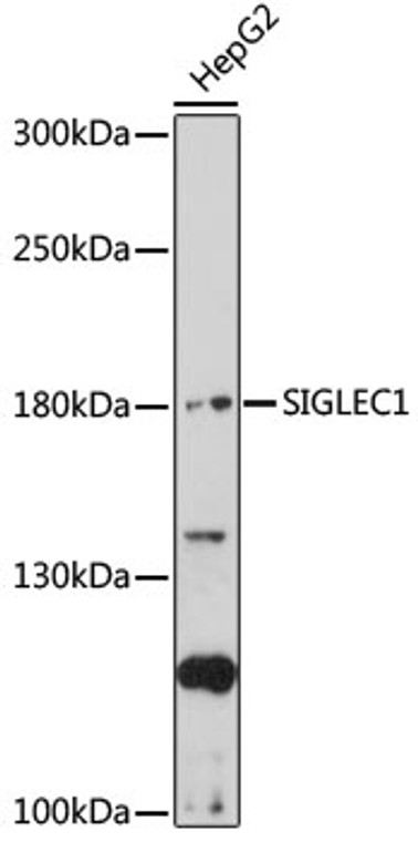
| Host: |
Rabbit |
| Applications: |
WB |
| Reactivity: |
Human |
| Note: |
STRICTLY FOR FURTHER SCIENTIFIC RESEARCH USE ONLY (RUO). MUST NOT TO BE USED IN DIAGNOSTIC OR THERAPEUTIC APPLICATIONS. |
| Short Description: |
Rabbit polyclonal antibody anti-SIGLEC1 (50-300) is suitable for use in Western Blot research applications. |
| Clonality: |
Polyclonal |
| Conjugation: |
Unconjugated |
| Isotype: |
IgG |
| Formulation: |
PBS with 0.01% Thimerosal, 50% Glycerol, pH7.3. |
| Purification: |
Affinity purification |
| Dilution Range: |
WB 1:500-1:2000 |
| Storage Instruction: |
Store at-20°C for up to 1 year from the date of receipt, and avoid repeat freeze-thaw cycles. |
| Gene Symbol: |
SIGLEC1 |
| Gene ID: |
6614 |
| Uniprot ID: |
SN_HUMAN |
| Immunogen Region: |
50-300 |
| Immunogen: |
Recombinant fusion protein containing a sequence corresponding to amino acids 50-300 of human SIGLEC1 (NP_075556.1). |
| Immunogen Sequence: |
EVPDGITAIWYYDYSGQRQV VSHSADPKLVEARFRGRTEF MGNPEHRVCNLLLKDLQPED SGSYNFRFEISEVNRWSDVK GTLVTVTEEPRVPTIASPVE LLEGTEVDFNCSTPYVCLQE QVRLQWQGQDPARSVTFNSQ KFEPTGVGHLETLHMAMSWQ DHGRILRCQLSVANHRAQSE IHLQVKYAPKGVKILLSPSG RNILPGELVTLTCQVNSSYP AVSSIKWLKDGVRLQTKTG |
| Tissue Specificity | Expressed by macrophages in various tissues. High levels are found in spleen, lymph node, perivascular macrophages in brain and lower levels in bone marrow, liver Kupffer cells and lamina propria of colon and lung. Also expressed by inflammatory macrophages in rheumatoid arthritis. |
| Function | Macrophage-restricted adhesion molecule that mediates sialic-acid dependent binding to lymphocytes, including granulocytes, monocytes, natural killer cells, B-cells and CD8 T-cells. Plays a crucial role in limiting bacterial dissemination by engaging sialylated bacteria to promote effective phagocytosis and antigen presentation for the adaptive immune response. Mediates the uptake of various enveloped viruses via sialic acid recognition and subsequently induces the formation of intracellular compartments filled with virions (VCCs). In turn, enhances macrophage-to-T-cell transmission of several viruses including HIV-1 or SARS-CoV-2. Acts as an endocytic receptor mediating clathrin dependent endocytosis. Preferentially binds to alpha-2,3-linked sialic acid. Binds to SPN/CD43 on T-cells. May play a role in hemopoiesis. Plays a role in the inhibition of antiviral innate immune by promoting TBK1 degradation via TYROBP and TRIM27-mediated ubiquitination. (Microbial infection) Facilitates viral cytoplasmic entry into activated dendritic cells via recognition of sialylated gangliosides pesent on viral membrane. |
| Protein Name | SialoadhesinSialic Acid-Binding Ig-Like Lectin 1Siglec-1Cd Antigen Cd169 |
| Database Links | Reactome: R-HSA-198933 |
| Cellular Localisation | Isoform 1: Cell MembraneSingle-Pass Type I Membrane ProteinIsoform 2: Secreted |
| Alternative Antibody Names | Anti-Sialoadhesin antibodyAnti-Sialic Acid-Binding Ig-Like Lectin 1 antibodyAnti-Siglec-1 antibodyAnti-Cd Antigen Cd169 antibodyAnti-SIGLEC1 antibodyAnti-SN antibody |
Information sourced from Uniprot.org
12 months for antibodies. 6 months for ELISA Kits. Please see website T&Cs for further guidance


