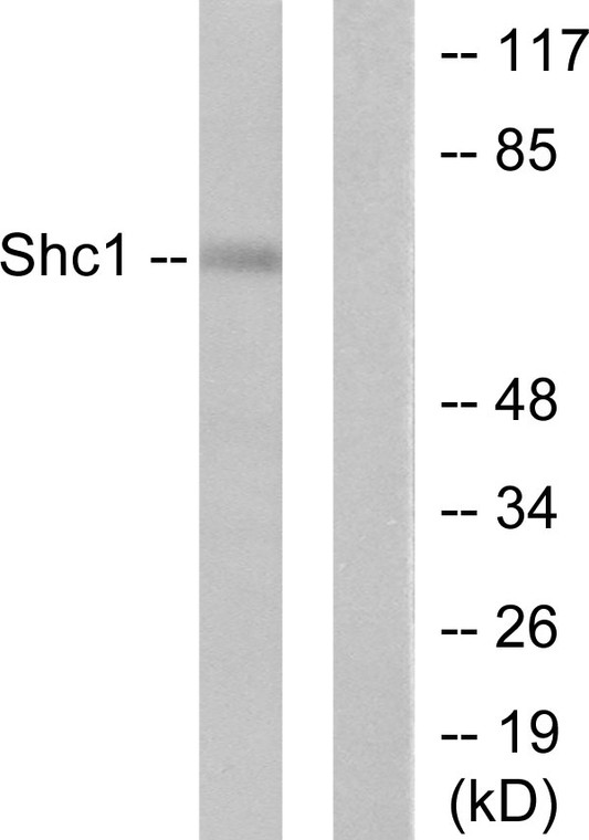
| Host: | Rabbit |
| Applications: | WB/IHC/IF/ELISA |
| Reactivity: | Human/Mouse/Rat |
| Note: | STRICTLY FOR FURTHER SCIENTIFIC RESEARCH USE ONLY (RUO). MUST NOT TO BE USED IN DIAGNOSTIC OR THERAPEUTIC APPLICATIONS. |
| Short Description: | Rabbit polyclonal antibody anti-SHC-transforming protein 1 (393-442 aa) is suitable for use in Western Blot, Immunohistochemistry, Immunofluorescence and ELISA research applications. |
| Clonality: | Polyclonal |
| Conjugation: | Unconjugated |
| Isotype: | IgG |
| Formulation: | Liquid in PBS containing 50% Glycerol, 0.5% BSA and 0.02% Sodium Azide. |
| Purification: | The antibody was affinity-purified from rabbit antiserum by affinity-chromatography using epitope-specific immunogen. |
| Concentration: | 1 mg/mL |
| Dilution Range: | WB 1:500-1:2000IHC 1:100-1:300ELISA 1:20000IF 1:50-200 |
| Storage Instruction: | Store at-20°C for up to 1 year from the date of receipt, and avoid repeat freeze-thaw cycles. |
| Gene Symbol: | SHC1 |
| Gene ID: | 6464 |
| Uniprot ID: | SHC1_HUMAN |
| Immunogen Region: | 393-442 aa |
| Specificity: | Shc Polyclonal Antibody detects endogenous levels of Shc protein. |
| Immunogen: | The antiserum was produced against synthesized peptide derived from the human Shc at the amino acid range 393-442 |
| Post Translational Modifications | Phosphorylated by activated epidermal growth factor receptor. Phosphorylated in response to FLT4 and KIT signaling. Isoform p46Shc and isoform p52Shc are phosphorylated on tyrosine residues of the Pro-rich domain. Isoform p66Shc is phosphorylated on Ser-36 by PRKCB upon treatment with insulin, hydrogen peroxide or irradiation with ultraviolet light. Tyrosine phosphorylated in response to FLT3 signaling. Tyrosine phosphorylated by activated PTK2B/PYK2. Tyrosine phosphorylated by ligand-activated ALK. Tyrosine phosphorylated by ligand-activated PDGFRB. Tyrosine phosphorylated by TEK/TIE2. May be tyrosine phosphorylated by activated PTK2/FAK1.tyrosine phosphorylation was seen in an astrocytoma biopsy, where PTK2/FAK1 kinase activity is high, but not in normal brain tissue. Isoform p52Shc dephosphorylation by PTPN2 may regulate interaction with GRB2. |
| Function | Signaling adapter that couples activated growth factor receptors to signaling pathways. Participates in a signaling cascade initiated by activated KIT and KITLG/SCF. Isoform p46Shc and isoform p52Shc, once phosphorylated, couple activated receptor tyrosine kinases to Ras via the recruitment of the GRB2/SOS complex and are implicated in the cytoplasmic propagation of mitogenic signals. Isoform p46Shc and isoform p52Shc may thus function as initiators of the Ras signaling cascade in various non-neuronal systems. Isoform p66Shc does not mediate Ras activation, but is involved in signal transduction pathways that regulate the cellular response to oxidative stress and life span. Isoform p66Shc acts as a downstream target of the tumor suppressor p53 and is indispensable for the ability of stress-activated p53 to induce elevation of intracellular oxidants, cytochrome c release and apoptosis. The expression of isoform p66Shc has been correlated with life span. Participates in signaling downstream of the angiopoietin receptor TEK/TIE2, and plays a role in the regulation of endothelial cell migration and sprouting angiogenesis. |
| Protein Name | Shc-Transforming Protein 1Shc-Transforming Protein 3Shc-Transforming Protein ASrc Homology 2 Domain-Containing-Transforming Protein C1Sh2 Domain Protein C1 |
| Database Links | Reactome: R-HSA-1236382Reactome: R-HSA-1250196Reactome: R-HSA-1250347Reactome: R-HSA-167044Reactome: R-HSA-180336Reactome: R-HSA-201556Reactome: R-HSA-210993Reactome: R-HSA-2424491 P29353-2Reactome: R-HSA-2428933Reactome: R-HSA-2730905 P29353-2Reactome: R-HSA-2871796 P29353-2Reactome: R-HSA-2871809 P29353-2Reactome: R-HSA-354192Reactome: R-HSA-381038Reactome: R-HSA-512988Reactome: R-HSA-5637810Reactome: R-HSA-5654688Reactome: R-HSA-5654699Reactome: R-HSA-5654704Reactome: R-HSA-5654719Reactome: R-HSA-5673001Reactome: R-HSA-74749Reactome: R-HSA-74751Reactome: R-HSA-8851805 P29353-2Reactome: R-HSA-8853659Reactome: R-HSA-8983432Reactome: R-HSA-9009391Reactome: R-HSA-9020558Reactome: R-HSA-9026519Reactome: R-HSA-9027284Reactome: R-HSA-9034864Reactome: R-HSA-912526Reactome: R-HSA-9634285Reactome: R-HSA-9634597Reactome: R-HSA-9664565Reactome: R-HSA-9665348Reactome: R-HSA-9665686Reactome: R-HSA-9674555Reactome: R-HSA-9680350Reactome: R-HSA-9725370Reactome: R-HSA-9842663 |
| Cellular Localisation | CytoplasmCell JunctionFocal AdhesionIsoform P46shc: Mitochondrion MatrixLocalized To The Mitochondria MatrixTargeting Of Isoform P46shc To Mitochondria Is Mediated By Its First 32 Amino AcidsWhich Behave As A Bona Fide Mitochondrial Targeting SequenceIsoform P52shc And Isoform P66shcThat Contain The Same Sequence But More Internally LocatedDisplay A Different Subcellular LocalizationIsoform P66shc: MitochondrionIn Case Of Oxidative ConditionsPhosphorylation At 'Ser-36' Of Isoform P66shcLeads To Mitochondrial Accumulation |
| Alternative Antibody Names | Anti-Shc-Transforming Protein 1 antibodyAnti-Shc-Transforming Protein 3 antibodyAnti-Shc-Transforming Protein A antibodyAnti-Src Homology 2 Domain-Containing-Transforming Protein C1 antibodyAnti-Sh2 Domain Protein C1 antibodyAnti-SHC1 antibodyAnti-SHC antibodyAnti-SHCA antibody |
Information sourced from Uniprot.org
12 months for antibodies. 6 months for ELISA Kits. Please see website T&Cs for further guidance


