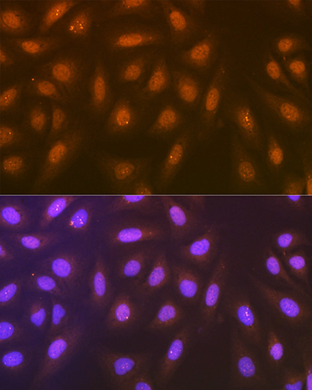
| Host: |
Rabbit |
| Applications: |
WB/IHC/IF |
| Reactivity: |
Human/Mouse/Rat |
| Note: |
STRICTLY FOR FURTHER SCIENTIFIC RESEARCH USE ONLY (RUO). MUST NOT TO BE USED IN DIAGNOSTIC OR THERAPEUTIC APPLICATIONS. |
| Short Description: |
Rabbit monoclonal antibody anti-Syntenin (1-100) is suitable for use in Western Blot, Immunohistochemistry and Immunofluorescence research applications. |
| Clonality: |
Monoclonal |
| Clone ID: |
S9MR |
| Conjugation: |
Unconjugated |
| Isotype: |
IgG |
| Formulation: |
PBS with 0.02% Sodium Azide, 0.05% BSA, 50% Glycerol, pH7.3. |
| Purification: |
Affinity purification |
| Dilution Range: |
WB 1:500-1:2000IHC-P 1:50-1:200IF/ICC 1:50-1:200 |
| Storage Instruction: |
Store at-20°C for up to 1 year from the date of receipt, and avoid repeat freeze-thaw cycles. |
| Gene Symbol: |
SDCBP |
| Gene ID: |
6386 |
| Uniprot ID: |
SDCB1_HUMAN |
| Immunogen Region: |
1-100 |
| Immunogen: |
A synthetic peptide corresponding to a sequence within amino acids 1-100 of human Syntenin (O00560). |
| Immunogen Sequence: |
MSLYPSLEDLKVDKVIQAQT AFSANPANPAILSEASAPIP HDGNLYPRLYPELSQYMGLS LNEEEIRANVAVVSGAPLQG QLVARPSSINYMVAPVTGND |
| Tissue Specificity | Expressed in lung cancers, including adenocarcinoma, squamous cell carcinoma and small-cell carcinoma (at protein level). Widely expressed. Expressed in fetal kidney, liver, lung and brain. In adult highest expression in heart and placenta. |
| Post Translational Modifications | Phosphorylated on tyrosine residues. |
| Function | Multifunctional adapter protein involved in diverse array of functions including trafficking of transmembrane proteins, neuro and immunomodulation, exosome biogenesis, and tumorigenesis. Positively regulates TGFB1-mediated SMAD2/3 activation and TGFB1-induced epithelial-to-mesenchymal transition (EMT) and cell migration in various cell types. May increase TGFB1 signaling by enhancing cell-surface expression of TGFR1 by preventing the interaction between TGFR1 and CAV1 and subsequent CAV1-dependent internalization and degradation of TGFR1. In concert with SDC1/4 and PDCD6IP, regulates exosome biogenesis. Regulates migration, growth, proliferation, and cell cycle progression in a variety of cancer types. In adherens junctions may function to couple syndecans to cytoskeletal proteins or signaling components. Seems to couple transcription factor SOX4 to the IL-5 receptor (IL5RA). May also play a role in vesicular trafficking. Seems to be required for the targeting of TGFA to the cell surface in the early secretory pathway. |
| Protein Name | Syntenin-1Melanoma Differentiation-Associated Protein 9Mda-9Pro-Tgf-Alpha Cytoplasmic Domain-Interacting Protein 18Tacip18Scaffold Protein Pbp1Syndecan-Binding Protein 1 |
| Database Links | Reactome: R-HSA-3928664Reactome: R-HSA-447043Reactome: R-HSA-5213460Reactome: R-HSA-5675482Reactome: R-HSA-6798695 |
| Cellular Localisation | Cell JunctionFocal AdhesionAdherens JunctionCell MembranePeripheral Membrane ProteinEndoplasmic Reticulum MembraneNucleusMelanosomeCytoplasmCytosolCytoskeletonSecretedExtracellular ExosomeMembrane RaftMainly Membrane-AssociatedLocalized To Adherens JunctionsFocal Adhesions And Endoplasmic ReticulumColocalized With Actin Stress FibersAlso Found In The NucleusIdentified By Mass Spectrometry In Melanosome Fractions From Stage I To Stage IvAssociated To The Plasma Membrane In The Presence Of Fzd7 And Phosphatidylinositol 45-Bisphosphate (Pip2) |
| Alternative Antibody Names | Anti-Syntenin-1 antibodyAnti-Melanoma Differentiation-Associated Protein 9 antibodyAnti-Mda-9 antibodyAnti-Pro-Tgf-Alpha Cytoplasmic Domain-Interacting Protein 18 antibodyAnti-Tacip18 antibodyAnti-Scaffold Protein Pbp1 antibodyAnti-Syndecan-Binding Protein 1 antibodyAnti-SDCBP antibodyAnti-MDA9 antibodyAnti-SYCL antibody |
Information sourced from Uniprot.org
12 months for antibodies. 6 months for ELISA Kits. Please see website T&Cs for further guidance


