| Host: |
Rabbit |
| Applications: |
WB |
| Reactivity: |
Mouse/Rat |
| Note: |
STRICTLY FOR FURTHER SCIENTIFIC RESEARCH USE ONLY (RUO). MUST NOT TO BE USED IN DIAGNOSTIC OR THERAPEUTIC APPLICATIONS. |
| Short Description: |
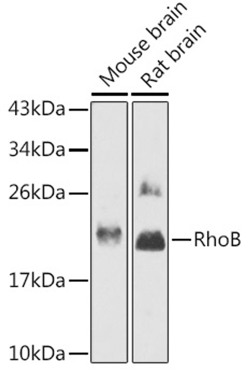
Rabbit polyclonal antibody anti-RHOB (1-196) is suitable for use in Western Blot research applications. |
| Clonality: |
Polyclonal |
| Conjugation: |
Unconjugated |
| Isotype: |
IgG |
| Formulation: |
PBS with 0.02% Sodium Azide, 50% Glycerol, pH7.3. |
| Purification: |
Affinity purification |
| Dilution Range: |
WB 1:500-1:2000 |
| Storage Instruction: |
Store at-20°C for up to 1 year from the date of receipt, and avoid repeat freeze-thaw cycles. |
| Gene Symbol: |
RHOB |
| Gene ID: |
388 |
| Uniprot ID: |
RHOB_HUMAN |
| Immunogen Region: |
1-196 |
| Immunogen: |
Recombinant fusion protein containing a sequence corresponding to amino acids 1-196 of human RhoB (NP_004031.1). |
| Immunogen Sequence: |
MAAIRKKLVVVGDGACGKTC LLIVFSKDEFPEVYVPTVFE NYVADIEVDGKQVELALWDT AGQEDYDRLRPLSYPDTDVI LMCFSVDSPDSLENIPEKWV PEVKHFCPNVPIILVANKKD LRSDEHVRTELARMKQEPVR TDDGRAMAVRIQAYDYLECS AKTKEGVREVFETATRAALQ KRYGSQNGCINCCKVL |
| Post Translational Modifications | Prenylation specifies the subcellular location of RHOB. The farnesylated form is localized to the plasma membrane while the geranylgeranylated form is localized to the endosome. (Microbial infection) Glycosylated at Tyr-34 by Photorhabdus asymbiotica toxin PAU_02230. Mono-O-GlcNAcylation by PAU_02230 inhibits downstream signaling by an impaired interaction with diverse regulator and effector proteins of Rho and leads to actin disassembly. (Microbial infection) Glucosylated at Thr-37 by C.difficile toxins TcdA and TcdB in the colonic epithelium. Monoglucosylation completely prevents the recognition of the downstream effector, blocking the GTPases in their inactive form, leading to actin cytoskeleton disruption. |
| Function | Mediates apoptosis in neoplastically transformed cells after DNA damage. Not essential for development but affects cell adhesion and growth factor signaling in transformed cells. Plays a negative role in tumorigenesis as deletion causes tumor formation. Involved in intracellular protein trafficking of a number of proteins. Targets PKN1 to endosomes and is involved in trafficking of the EGF receptor from late endosomes to lysosomes. Also required for stability and nuclear trafficking of AKT1/AKT which promotes endothelial cell survival during vascular development. Serves as a microtubule-dependent signal that is required for the myosin contractile ring formation during cell cycle cytokinesis. Required for genotoxic stress-induced cell death in breast cancer cells. |
| Protein Name | Rho-Related Gtp-Binding Protein RhobRho Cdna Clone 6H6 |
| Database Links | Reactome: R-HSA-114604Reactome: R-HSA-416482Reactome: R-HSA-416572Reactome: R-HSA-5625740Reactome: R-HSA-5625900Reactome: R-HSA-5627117Reactome: R-HSA-5663220Reactome: R-HSA-5666185Reactome: R-HSA-9013026 |
| Cellular Localisation | Late Endosome MembraneLipid-AnchorCell MembraneNucleusCleavage FurrowLate Endosomal Membrane (Geranylgeranylated Form)Plasma Membrane (Farnesylated Form)Also Detected At The Nuclear Margin And In The NucleusTranslocates To The Equatorial Region Before Furrow Formation In A Ect2-Dependent Manner |
| Alternative Antibody Names | Anti-Rho-Related Gtp-Binding Protein Rhob antibodyAnti-Rho Cdna Clone 6 antibodyAnti-H6 antibodyAnti-RHOB antibodyAnti-ARH6 antibodyAnti-ARHB antibody |
Information sourced from Uniprot.org
12 months for antibodies. 6 months for ELISA Kits. Please see website T&Cs for further guidance


