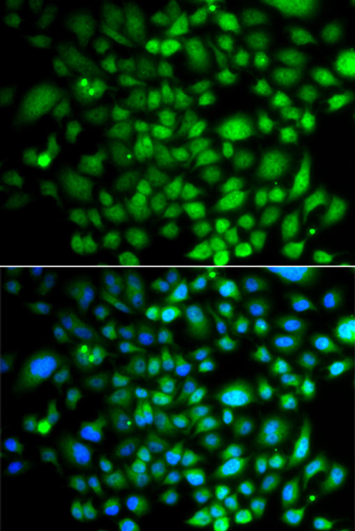
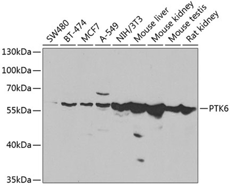
| Host: |
Rabbit |
| Applications: |
WB/IF |
| Reactivity: |
Human/Mouse/Rat |
| Note: |
STRICTLY FOR FURTHER SCIENTIFIC RESEARCH USE ONLY (RUO). MUST NOT TO BE USED IN DIAGNOSTIC OR THERAPEUTIC APPLICATIONS. |
| Short Description: |
Rabbit polyclonal antibody anti-PTK6 (222-451) is suitable for use in Western Blot and Immunofluorescence research applications. |
| Clonality: |
Polyclonal |
| Conjugation: |
Unconjugated |
| Isotype: |
IgG |
| Formulation: |
PBS with 0.02% Sodium Azide, 50% Glycerol, pH7.3. |
| Purification: |
Affinity purification |
| Dilution Range: |
WB 1:500-1:2000IF/ICC 1:50-1:200 |
| Storage Instruction: |
Store at-20°C for up to 1 year from the date of receipt, and avoid repeat freeze-thaw cycles. |
| Gene Symbol: |
PTK6 |
| Gene ID: |
5753 |
| Uniprot ID: |
PTK6_HUMAN |
| Immunogen Region: |
222-451 |
| Immunogen: |
Recombinant fusion protein containing a sequence corresponding to amino acids 222-451 of human PTK6 (NP_005966.1). |
| Immunogen Sequence: |
SRDNLLHQQMLQSEIQAMKK LRHKHILALYAVVSVGDPVY IITELMAKGSLLELLRDSDE KVLPVSELLDIAWQVAEGMC YLESQNYIHRDLAARNILVG ENTLCKVGDFGLARLIKEDV YLSHDHNIPYKWTAPEALSR GHYSTKSDVWSFGILLHEMF SRGQVPYPGMSNHEAFLRVD AGYRMPCPLECPPSVHKLML TCWCRDPEQRPCFKALRERL SSFTSYENPT |
| Tissue Specificity | Epithelia-specific. Very high level in colon and high levels in small intestine and prostate, and low levels in some fetal tissues. Not expressed in breast or ovarian tissue but expressed in high percentage of breast and ovarian cancers. Also overexpressed in some metastatic melanomas, lymphomas, colon cancers, squamous cell carcinomas and prostate cancers. Also found in melanocytes. Not expressed in heart, brain, placenta, lung, liver, skeletal muscle, kidney and pancreas. Isoform 2 is present in prostate epithelial cell lines derived from normal prostate and prostate adenocarcinomas, as well as in a variety of cell lines. |
| Post Translational Modifications | Autophosphorylated. Autophosphorylation of Tyr-342 leads to an increase of kinase activity. Tyr-447 binds to the SH2 domain when phosphorylated and negatively regulates kinase activity. |
| Function | Non-receptor tyrosine-protein kinase implicated in the regulation of a variety of signaling pathways that control the differentiation and maintenance of normal epithelia, as well as tumor growth. Function seems to be context dependent and differ depending on cell type, as well as its intracellular localization. A number of potential nuclear and cytoplasmic substrates have been identified. These include the RNA-binding proteins: KHDRBS1/SAM68, KHDRBS2/SLM1, KHDRBS3/SLM2 and SFPQ/PSF.transcription factors: STAT3 and STAT5A/B and a variety of signaling molecules: ARHGAP35/p190RhoGAP, PXN/paxillin, BTK/ATK, STAP2/BKS. Associates also with a variety of proteins that are likely upstream of PTK6 in various signaling pathways, or for which PTK6 may play an adapter-like role. These proteins include ADAM15, EGFR, ERBB2, ERBB3 and IRS4. In normal or non-tumorigenic tissues, PTK6 promotes cellular differentiation and apoptosis. In tumors PTK6 contributes to cancer progression by sensitizing cells to mitogenic signals and enhancing proliferation, anchorage-independent survival and migration/invasion. Association with EGFR, ERBB2, ERBB3 may contribute to mammary tumor development and growth through enhancement of EGF-induced signaling via BTK/AKT and PI3 kinase. Contributes to migration and proliferation by contributing to EGF-mediated phosphorylation of ARHGAP35/p190RhoGAP, which promotes association with RASA1/p120RasGAP, inactivating RhoA while activating RAS. EGF stimulation resulted in phosphorylation of PNX/Paxillin by PTK6 and activation of RAC1 via CRK/CrKII, thereby promoting migration and invasion. PTK6 activates STAT3 and STAT5B to promote proliferation. Nuclear PTK6 may be important for regulating growth in normal epithelia, while cytoplasmic PTK6 might activate oncogenic signaling pathways. Isoform 2 inhibits PTK6 phosphorylation and PTK6 association with other tyrosine-phosphorylated proteins. |
| Protein Name | Protein-Tyrosine Kinase 6Breast Tumor KinaseTyrosine-Protein Kinase Brk |
| Database Links | Reactome: R-HSA-187577Reactome: R-HSA-69231Reactome: R-HSA-8847993Reactome: R-HSA-8849468Reactome: R-HSA-8849469Reactome: R-HSA-8849470Reactome: R-HSA-8849471Reactome: R-HSA-8849472Reactome: R-HSA-8849473Reactome: R-HSA-8849474Reactome: R-HSA-8857538Reactome: R-HSA-9707564 |
| Cellular Localisation | CytoplasmNucleusCell ProjectionRuffleMembraneColocalizes With Khdrbs1Khdrbs2 Or Khdrbs3Within The NucleusNuclear Localization In Epithelial Cells Of Normal Prostate But Cytoplasmic Localization In Cancer Prostate |
| Alternative Antibody Names | Anti-Protein-Tyrosine Kinase 6 antibodyAnti-Breast Tumor Kinase antibodyAnti-Tyrosine-Protein Kinase Brk antibodyAnti-PTK6 antibodyAnti-BRK antibody |
Information sourced from Uniprot.org
12 months for antibodies. 6 months for ELISA Kits. Please see website T&Cs for further guidance


