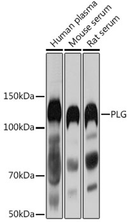
| Host: |
Rabbit |
| Applications: |
WB |
| Reactivity: |
Human/Mouse/Rat |
| Note: |
STRICTLY FOR FURTHER SCIENTIFIC RESEARCH USE ONLY (RUO). MUST NOT TO BE USED IN DIAGNOSTIC OR THERAPEUTIC APPLICATIONS. |
| Short Description: |
Rabbit polyclonal antibody anti-PLG (20-300) is suitable for use in Western Blot research applications. |
| Clonality: |
Polyclonal |
| Conjugation: |
Unconjugated |
| Isotype: |
IgG |
| Formulation: |
PBS with 0.02% Sodium Azide, 50% Glycerol, pH7.3. |
| Purification: |
Affinity purification |
| Dilution Range: |
WB 1:500-1:2000 |
| Storage Instruction: |
Store at-20°C for up to 1 year from the date of receipt, and avoid repeat freeze-thaw cycles. |
| Gene Symbol: |
PLG |
| Gene ID: |
5340 |
| Uniprot ID: |
PLMN_HUMAN |
| Immunogen Region: |
20-300 |
| Immunogen: |
Recombinant fusion protein containing a sequence corresponding to amino acids 20-300 of human PLG (NP_000292.1). |
| Immunogen Sequence: |
EPLDDYVNTQGASLFSVTKK QLGAGSIEECAAKCEEDEEF TCRAFQYHSKEQQCVIMAEN RKSSIIIRMRDVVLFEKKVY LSECKTGNGKNYRGTMSKTK NGITCQKWSSTSPHRPRFSP ATHPSEGLEENYCRNPDNDP QGPWCYTTDPEKRYDYCDIL ECEEECMHCSGENYDGKISK TMSGLECQAWDSQSPHAHGY IPSKFPNKNLKKNYCRNPDR ELRPWCFTTDPNKRWELCD |
| Tissue Specificity | Present in plasma and many other extracellular fluids. It is synthesized in the liver. |
| Post Translational Modifications | N-linked glycan contains N-acetyllactosamine and sialic acid. O-linked glycans consist of Gal-GalNAc disaccharide modified with up to 2 sialic acid residues (microheterogeneity). In the presence of the inhibitor, the activation involves only cleavage after Arg-580, yielding two chains held together by two disulfide bonds. In the absence of the inhibitor, the activation involves additionally the removal of the activation peptide. |
| Function | Plasmin dissolves the fibrin of blood clots and acts as a proteolytic factor in a variety of other processes including embryonic development, tissue remodeling, tumor invasion, and inflammation. In ovulation, weakens the walls of the Graafian follicle. It activates the urokinase-type plasminogen activator, collagenases and several complement zymogens, such as C1 and C5. Cleavage of fibronectin and laminin leads to cell detachment and apoptosis. Also cleaves fibrin, thrombospondin and von Willebrand factor. Its role in tissue remodeling and tumor invasion may be modulated by CSPG4. Binds to cells. Angiostatin is an angiogenesis inhibitor that blocks neovascularization and growth of experimental primary and metastatic tumors in vivo. (Microbial infection) ENO/enoloase from parasite P.falciparum (strain NF54) interacts with PLG present in the mosquito blood meal to promote the invasion of the mosquito midgut by the parasite ookinete. The catalytic active form, plasmin, is essential for the invasion of the mosquito midgut. (Microbial infection) Binds to OspC on the surface of B.burgdorferi cells, possibly conferring an extracellular protease activity on the bacteria that allows it to traverse host tissue. |
| Protein Name | Plasminogen Cleaved Into - Plasmin Heavy Chain A - Activation Peptide - Angiostatin - Plasmin Heavy Chain A - Short Form - Plasmin Light Chain B |
| Database Links | Reactome: R-HSA-114608Reactome: R-HSA-1474228Reactome: R-HSA-1592389Reactome: R-HSA-186797Reactome: R-HSA-381426Reactome: R-HSA-75205 |
| Cellular Localisation | SecretedLocates To The Cell Surface Where It Is Proteolytically Cleaved To Produce The Active PlasminInteraction With Hrg Tethers It To The Cell Surface |
| Alternative Antibody Names | Anti-Plasminogen Cleaved Into - Plasmin Heavy Chain A - Activation Peptide - Angiostatin - Plasmin Heavy Chain A - Short Form - Plasmin Light Chain B antibodyAnti-PLG antibody |
Information sourced from Uniprot.org
12 months for antibodies. 6 months for ELISA Kits. Please see website T&Cs for further guidance


