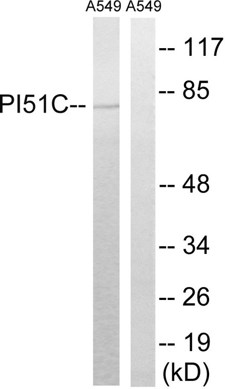
| Host: |
Rabbit |
| Applications: |
WB/ELISA |
| Reactivity: |
Human/Rat/Mouse |
| Note: |
STRICTLY FOR FURTHER SCIENTIFIC RESEARCH USE ONLY (RUO). MUST NOT TO BE USED IN DIAGNOSTIC OR THERAPEUTIC APPLICATIONS. |
| Short Description: |
Rabbit polyclonal antibody anti-Phosphatidylinositol 4-phosphate 5-kinase type-1 gammaP-5-kinase 1 gamma (305-354 aa) is suitable for use in Western Blot and ELISA research applications. |
| Clonality: |
Polyclonal |
| Conjugation: |
Unconjugated |
| Isotype: |
IgG |
| Formulation: |
Liquid in PBS containing 50% Glycerol, 0.5% BSA and 0.02% Sodium Azide. |
| Purification: |
The antibody was affinity-purified from rabbit antiserum by affinity-chromatography using epitope-specific immunogen. |
| Concentration: |
1 mg/mL |
| Dilution Range: |
WB 1:500-1:2000ELISA 1:20000 |
| Storage Instruction: |
Store at-20°C for up to 1 year from the date of receipt, and avoid repeat freeze-thaw cycles. |
| Gene Symbol: |
PIP5K1C |
| Gene ID: |
23396 |
| Uniprot ID: |
PI51C_HUMAN |
| Immunogen Region: |
305-354 aa |
| Specificity: |
PIPK I Gamma Polyclonal Antibody detects endogenous levels of PIPK I Gamma protein. |
| Immunogen: |
The antiserum was produced against synthesized peptide derived from the human PIP5K1C at the amino acid range 305-354 |
| Post Translational Modifications | Phosphorylation on Ser-650 negatively regulates binding to TLN2 and is strongly stimulated in mitosis. Phosphorylation on Tyr-649 is necessary for targeting to focal adhesions. Phosphorylation on Ser-650 and Tyr-649 are mutually exclusive. Phosphorylated by SYK and CSK. Tyrosine phosphorylation is enhanced by PTK2 signaling. Phosphorylated at Tyr-639 upon EGF stimulation. Some studies suggest that phosphorylation on Tyr-649 enhances binding to tailins (TLN1 and TLN2). phosphorylation at Tyr-649 does not directly enhance binding to tailins (TLN1 and TLN2) but may act indirectly by inhibiting phosphorylation at Ser-650. Acetylation at Lys-265 and Lys-268 seems to decrease lipid 1-phosphatidylinositol-4-phosphate 5-kinase activity. Deacetylation of these sites by SIRT1 positively regulates the exocytosis of TSH-containing granules from pituitary cells. |
| Function | Catalyzes the phosphorylation of phosphatidylinositol 4-phosphate (PtdIns(4)P/PI4P) to form phosphatidylinositol 4,5-bisphosphate (PtdIns(4,5)P2/PIP2), a lipid second messenger that regulates several cellular processes such as signal transduction, vesicle trafficking, actin cytoskeleton dynamics, cell adhesion, and cell motility. PtdIns(4,5)P2 can directly act as a second messenger or can be utilized as a precursor to generate other second messengers: inositol 1,4,5-trisphosphate (IP3), diacylglycerol (DAG) or phosphatidylinositol-3,4,5-trisphosphate (PtdIns(3,4,5)P3/PIP3) (Probable). PIP5K1A-mediated phosphorylation of PtdIns(4)P is the predominant pathway for PtdIns(4,5)P2 synthesis. Together with PIP5K1A, is required for phagocytosis, both enzymes regulating different types of actin remodeling at sequential steps. Promotes particle attachment by generating the pool of PtdIns(4,5)P2 that induces controlled actin depolymerization to facilitate Fc-gamma-R clustering. Mediates RAC1-dependent reorganization of actin filaments. Required for synaptic vesicle transport. Controls the plasma membrane pool of PtdIns(4,5)P2 implicated in synaptic vesicle endocytosis and exocytosis. Plays a role in endocytosis mediated by clathrin and AP-2 (adaptor protein complex 2). Required for clathrin-coated pits assembly at the synapse. Participates in cell junction assembly. Modulates adherens junctions formation by facilitating CDH1/cadherin trafficking. Required for focal adhesion dynamics. Modulates the targeting of talins (TLN1 and TLN2) to the plasma membrane and their efficient assembly into focal adhesions. Regulates the interaction between talins (TLN1 and TLN2) and beta-integrins. Required for uropodium formation and retraction of the cell rear during directed migration. Has a role in growth factor-stimulated directional cell migration and adhesion. Required for talin assembly into nascent adhesions forming at the leading edge toward the direction of the growth factor. Negative regulator of T-cell activation and adhesion. Negatively regulates integrin alpha-L/beta-2 (LFA-1) polarization and adhesion induced by T-cell receptor. Together with PIP5K1A has a role during embryogenesis and together with PIP5K1B may have a role immediately after birth. |
| Protein Name | Phosphatidylinositol 4-Phosphate 5-Kinase Type-1 GammaPip5k1gammaPtdins(4p-5-Kinase 1 GammaType I Phosphatidylinositol 4-Phosphate 5-Kinase Gamma |
| Database Links | Reactome: R-HSA-1660499Reactome: R-HSA-399955Reactome: R-HSA-6811558Reactome: R-HSA-8856828 |
| Cellular Localisation | Cell MembranePeripheral Membrane ProteinCytoplasmic SideEndomembrane SystemCytoplasmCell JunctionFocal AdhesionAdherens JunctionCell ProjectionRuffle MembranePhagocytic CupUropodiumDetected In Plasma Membrane InvaginationsIsoform 3 Is Detected In Intracellular Vesicle-Like StructuresIsoform 2: CytoplasmNucleus |
| Alternative Antibody Names | Anti-Phosphatidylinositol 4-Phosphate 5-Kinase Type-1 Gamma antibodyAnti-Pip5k1gamma antibodyAnti-Ptdins(4p-5-Kinase 1 Gamma antibodyAnti-Type I Phosphatidylinositol 4-Phosphate 5-Kinase Gamma antibodyAnti-PIP5K1C antibodyAnti-KIAA0589 antibody |
Information sourced from Uniprot.org
12 months for antibodies. 6 months for ELISA Kits. Please see website T&Cs for further guidance


