| Host: |
Rabbit |
| Applications: |
WB/IHC/IF/ELISA |
| Reactivity: |
Human/Rat/Mouse |
| Note: |
STRICTLY FOR FURTHER SCIENTIFIC RESEARCH USE ONLY (RUO). MUST NOT TO BE USED IN DIAGNOSTIC OR THERAPEUTIC APPLICATIONS. |
| Short Description: |
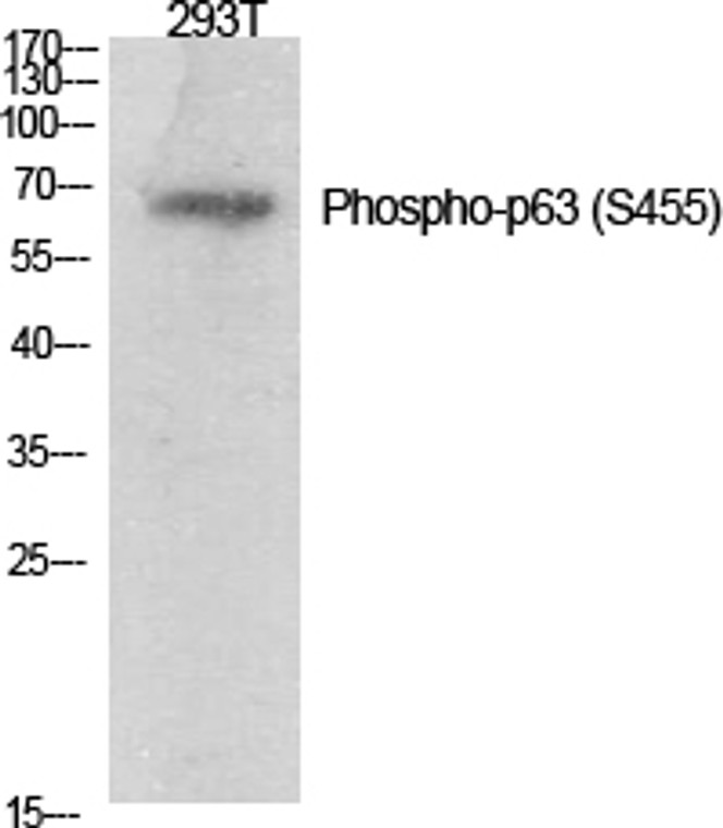
Rabbit polyclonal antibody anti-Phospho-Tumor protein 63-Ser455 (421-470 aa) is suitable for use in Western Blot, Immunohistochemistry, Immunofluorescence and ELISA research applications. |
| Clonality: |
Polyclonal |
| Conjugation: |
Unconjugated |
| Isotype: |
IgG |
| Formulation: |
Liquid in PBS containing 50% Glycerol, 0.5% BSA and 0.02% Sodium Azide. |
| Purification: |
The antibody was affinity-purified from rabbit antiserum by affinity-chromatography using epitope-specific immunogen. |
| Concentration: |
1 mg/mL |
| Dilution Range: |
WB 1:500-1:2000IHC 1:100-1:300ELISA 1:20000IF 1:50-200 |
| Storage Instruction: |
Store at-20°C for up to 1 year from the date of receipt, and avoid repeat freeze-thaw cycles. |
| Gene Symbol: |
TP63 |
| Gene ID: |
8626 |
| Uniprot ID: |
P63_HUMAN |
| Immunogen Region: |
421-470 aa |
| Specificity: |
Phospho-p63 (S455) Polyclonal Antibody detects endogenous levels of p63 protein only when phosphorylated at S455. |
| Immunogen: |
The antiserum was produced against synthesized peptide derived from the human p63 around the phosphorylation site of Ser455 at the amino acid range 421-470 |
| Post Translational Modifications | May be sumoylated. Ubiquitinated. Polyubiquitination involves WWP1 and leads to proteasomal degradation of this protein. |
| Function | Acts as a sequence specific DNA binding transcriptional activator or repressor. The isoforms contain a varying set of transactivation and auto-regulating transactivation inhibiting domains thus showing an isoform specific activity. Isoform 2 activates RIPK4 transcription. May be required in conjunction with TP73/p73 for initiation of p53/TP53 dependent apoptosis in response to genotoxic insults and the presence of activated oncogenes. Involved in Notch signaling by probably inducing JAG1 and JAG2. Plays a role in the regulation of epithelial morphogenesis. The ratio of DeltaN-type and TA*-type isoforms may govern the maintenance of epithelial stem cell compartments and regulate the initiation of epithelial stratification from the undifferentiated embryonal ectoderm. Required for limb formation from the apical ectodermal ridge. Activates transcription of the p21 promoter. |
| Protein Name | Tumor Protein 63P63Chronic Ulcerative Stomatitis ProteinCuspKeratinocyte Transcription Factor KetTransformation-Related Protein 63Tp63Tumor Protein P73-LikeP73lP40P51 |
| Database Links | Reactome: R-HSA-139915Reactome: R-HSA-5620971Reactome: R-HSA-5628897Reactome: R-HSA-6803204Reactome: R-HSA-6803205Reactome: R-HSA-6803207Reactome: R-HSA-6803211Reactome: R-HSA-6804759 |
| Cellular Localisation | Nucleus |
| Alternative Antibody Names | Anti-Tumor Protein 63 antibodyAnti-P63 antibodyAnti-Chronic Ulcerative Stomatitis Protein antibodyAnti-Cusp antibodyAnti-Keratinocyte Transcription Factor Ket antibodyAnti-Transformation-Related Protein 63 antibodyAnti-Tp63 antibodyAnti-Tumor Protein P73-Like antibodyAnti-P73l antibodyAnti-P40 antibodyAnti-P51 antibodyAnti-TP63 antibodyAnti-KET antibodyAnti-P63 antibodyAnti-P73H antibodyAnti-P73L antibodyAnti-TP73L antibody |
Information sourced from Uniprot.org
12 months for antibodies. 6 months for ELISA Kits. Please see website T&Cs for further guidance


