-
Western blot analysis of TF-1 cells, using Phospho-STAT5A-Y694 rabbit monoclonal antibody (STJ11102567) at 1:1000 dilution. TF-1 cells were treated by GM-CSF (25 ng/mL) at 37 °C for 30 minutes after serum-starvation overnight. Secondary antibody: HRP Goat Anti-rabbit IgG (H+L) (STJS000856) at 1:10000 dilution. Lysates/proteins: 25 Mu g per lane. Blocking buffer: 3% non-fat dry milk in TBST. Detection: ECL Basic Kit. Exposure time: 60s.
-
Immunohistochemistry analysis of paraffin-embedded rat brain using Phospho-STAT5A-Y694 antibody (STJ11102567) at dilution of 1:100 (40x lens). Perform microwave antigen retrieval with 10 mM Tris/EDTA buffer pH 9. 0 before commencing with immunohistochemistry staining protocol.
-
Immunohistochemistry analysis of paraffin-embedded human colon carcinoma using Phospho-STAT5A-Y694 rabbit monoclonal antibody (STJ11102567) at dilution of 1:200 (40x lens). Perform high pressure antigen retrieval with 10 mM citrate buffer pH 6. 0 before commencing with immunohistochemistry staining protocol.
-
Immunohistochemistry analysis of paraffin-embedded human colon using Phospho-STAT5A-Y694 rabbit monoclonal antibody (STJ11102567) at dilution of 1:200 (40x lens). Perform high pressure antigen retrieval with 10 mM citrate buffer pH 6. 0 before commencing with immunohistochemistry staining protocol.
-
Immunohistochemistry analysis of paraffin-embedded human kidney using Phospho-STAT5A-Y694 rabbit monoclonal antibody (STJ11102567) at dilution of 1:200 (40x lens). Perform high pressure antigen retrieval with 10 mM citrate buffer pH 6. 0 before commencing with immunohistochemistry staining protocol.
-
Immunohistochemistry analysis of paraffin-embedded human liver using Phospho-STAT5A-Y694 rabbit monoclonal antibody (STJ11102567) at dilution of 1:200 (40x lens). Perform high pressure antigen retrieval with 10 mM citrate buffer pH 6. 0 before commencing with immunohistochemistry staining protocol.
-
Immunohistochemistry analysis of paraffin-embedded human placenta using Phospho-STAT5A-Y694 rabbit monoclonal antibody (STJ11102567) at dilution of 1:200 (40x lens). Perform high pressure antigen retrieval with 10 mM citrate buffer pH 6. 0 before commencing with immunohistochemistry staining protocol.
-
Immunohistochemistry analysis of paraffin-embedded human spleen using Phospho-STAT5A-Y694 rabbit monoclonal antibody (STJ11102567) at dilution of 1:200 (40x lens). Perform high pressure antigen retrieval with 10 mM citrate buffer pH 6. 0 before commencing with immunohistochemistry staining protocol.
-
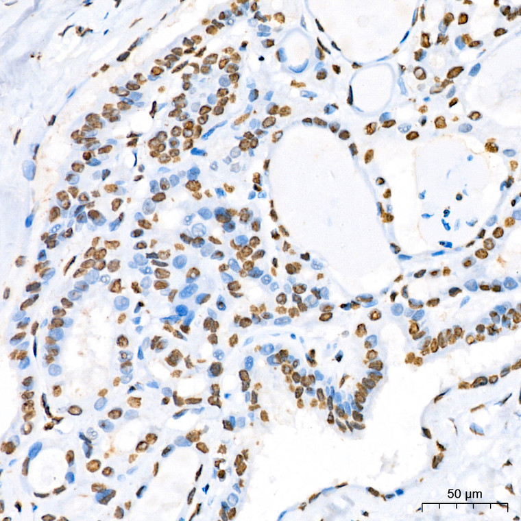
Immunohistochemistry analysis of paraffin-embedded human thyroid cancer using Phospho-STAT5A-Y694 rabbit monoclonal antibody (STJ11102567) at dilution of 1:200 (40x lens). Perform high pressure antigen retrieval with 10 mM citrate buffer pH 6. 0 before commencing with immunohistochemistry staining protocol.
-
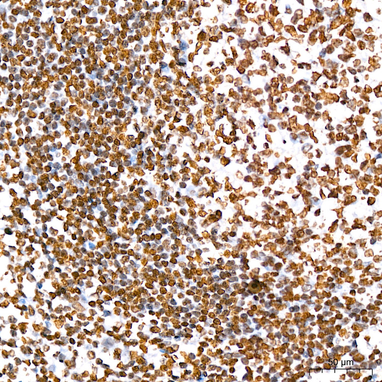
Immunohistochemistry analysis of paraffin-embedded human tonsil using Phospho-STAT5A-Y694 rabbit monoclonal antibody (STJ11102567) at dilution of 1:200 (40x lens). Perform high pressure antigen retrieval with 10 mM citrate buffer pH 6. 0 before commencing with immunohistochemistry staining protocol.
-
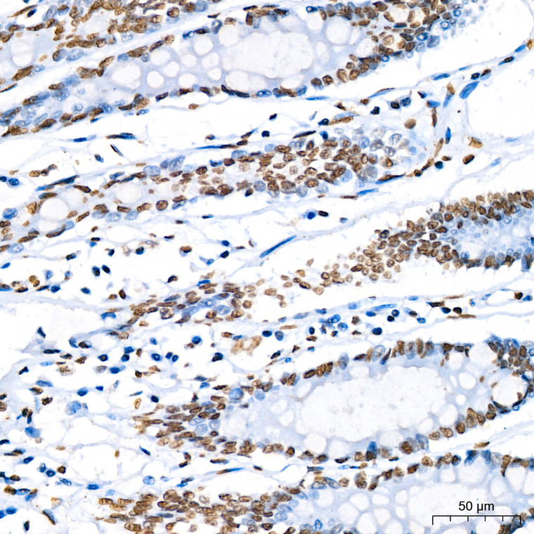
Immunohistochemistry analysis of paraffin-embedded mouse intestin using Phospho-STAT5A-Y694 rabbit monoclonal antibody (STJ11102567) at dilution of 1:200 (40x lens). Perform high pressure antigen retrieval with 10 mM citrate buffer pH 6. 0 before commencing with immunohistochemistry staining protocol.
-
Immunohistochemistry analysis of paraffin-embedded mouse liver using Phospho-STAT5A-Y694 rabbit monoclonal antibody (STJ11102567) at dilution of 1:200 (40x lens). Perform high pressure antigen retrieval with 10 mM citrate buffer pH 6. 0 before commencing with immunohistochemistry staining protocol.
-
Immunohistochemistry analysis of paraffin-embedded rat kidney using Phospho-STAT5A-Y694 rabbit monoclonal antibody (STJ11102567) at dilution of 1:200 (40x lens). Perform high pressure antigen retrieval with 10 mM citrate buffer pH 6. 0 before commencing with immunohistochemistry staining protocol.


