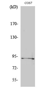
 Polyclonal Antibody diluted at 1:1000 STJ90408")
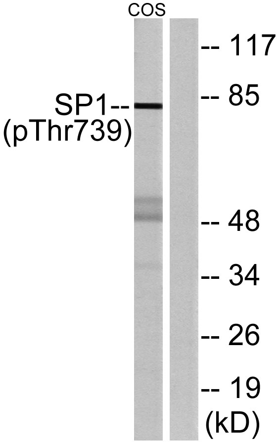
 Antibody. STJ90408")
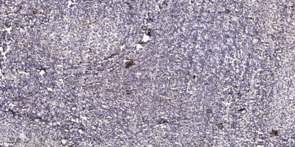
. 2, STJ90408")
 for Immunogen Phosphopeptide (Phospho-left) and Non-Phosphopeptide STJ90408")
 Polyclonal Antibody diluted at 1:1000 STJ90408")
Anti-Phospho-SP1-Thr739 antibody (706-755 aa) (STJ90408)
SPECIFICATIONS
ClonalityPolyclonal
HostRabbit
ConjugationUnconjugated
IsotypeIgG
ImmunogenThe antiserum was produced against synthesized peptide derived from the human SP1 around the phosphorylation site of Thr739 at the amino acid range 706-755
General Information
| Short Description | Rabbit polyclonal anti-Phospho-Transcription factor Sp1-Thr739 (706-755 aa) for use in WB, IHC, IF and ELISA in Human, Mouse, Rat and Monkey samples. Datasheet included with dilution recommendations, and related reagents. |
| Applications | WB/IHC/IF/ELISA |
| Host | Rabbit |
| Reactivity | Human/Mouse/Rat/Monkey |
| Note | STRICTLY FOR FURTHER SCIENTIFIC RESEARCH USE ONLY (RUO). MUST NOT TO BE USED IN DIAGNOSTIC OR THERAPEUTIC APPLICATIONS. |
Product Properties
| Clonality | Polyclonal |
| Isotype | IgG |
| Conjugation | Unconjugated |
| Concentration | 1 mg/mL |
| Purification | The antibody was affinity-purified from rabbit antiserum by affinity-chromatography using epitope-specific immunogen. |
| Dilution Range | WB 1:500-1:2000IHC 1:100-1:300ELISA 1:5000IF 1:50-200 |
| Formulation | Liquid in PBS containing 50% Glycerol, 0.5% BSA and 0.02% Sodium Azide. |
| Storage Instruction | Store at-20°C for up to 1 year from the date of receipt, and avoid repeat freeze-thaw cycles. |
Target Information
| Gene Symbol | SP1 |
| Gene ID | 6667 |
| Uniprot ID | SP1_HUMAN |
| Immunogen | The antiserum was produced against synthesized peptide derived from the human SP1 around the phosphorylation site of Thr739 at the amino acid range 706-755 |
| Immunogen Region | 706-755 aa |
| Specificity | Phospho-Sp1 (T739) Polyclonal Antibody detects endogenous levels of Sp1 protein only when phosphorylated at T739. |
Additional Info
| Post Translational Modifications | Phosphorylated on multiple serine and threonine residues. Phosphorylation is coupled to ubiquitination, sumoylation and proteolytic processing. Phosphorylation on Ser-59 enhances proteolytic cleavage. Phosphorylation on Ser-7 enhances ubiquitination and protein degradation. Hyperphosphorylation on Ser-101 in response to DNA damage has no effect on transcriptional activity. MAPK1/MAPK3-mediated phosphorylation on Thr-453 and Thr-739 enhances VEGF transcription but, represses FGF2-triggered PDGFR-alpha transcription. Also implicated in the repression of RECK by ERBB2. Hyperphosphorylated on Thr-278 and Thr-739 during mitosis by MAPK8 shielding SP1 from degradation by the ubiquitin-dependent pathway. Phosphorylated in the zinc-finger domain by calmodulin-activated PKCzeta. Phosphorylation on Ser-641 by PKCzeta is critical for TSA-activated LHR gene expression through release of its repressor, p107. Phosphorylation on Thr-668, Ser-670 and Thr-681 is stimulated by angiotensin II via the AT1 receptor inducing increased binding to the PDGF-D promoter. This phosphorylation is increased in injured artey wall. Ser-59 and Thr-681 can both be dephosphorylated by PP2A during cell-cycle interphase. Dephosphorylation on Ser-59 leads to increased chromatin association during interphase and increases the transcriptional activity. On insulin stimulation, sequentially glycosylated and phosphorylated on several C-terminal serine and threonine residues. Acetylated. Acetylation/deacetylation events affect transcriptional activity. Deacetylation leads to an increase in the expression of the 12(s)-lipooxygenase gene through recruitment of p300 to the promoter. Deacetylated by HDAC6 which leads to increased expression of ENG and positive regulation of angiogenesis. Ubiquitinated. Ubiquitination occurs on the C-terminal proteolytically-cleaved peptide and is triggered by phosphorylation. Sumoylated with SUMO1. Sumoylation modulates proteolytic cleavage of the N-terminal repressor domain. Sumoylation levels are attenuated during tumorigenesis. Phosphorylation mediates SP1 desumoylation. Proteolytic cleavage in the N-terminal repressor domain is prevented by sumoylation. The C-terminal cleaved product is susceptible to degradation. O-glycosylated.Contains 8 N-acetylglucosamine side chains. Levels are controlled by insulin and the SP1 phosphorylation states. Insulin-mediated O-glycosylation locates SP1 to the nucleus, where it is sequentially deglycosylated and phosphorylated. O-glycosylation affects transcriptional activity through disrupting the interaction with a number of transcription factors including ELF1 and NFYA. Also inhibits interaction with the HIV1 promoter. Inhibited by peroxisomome proliferator receptor gamma (PPARgamma). |
| Function | Transcription factor that can activate or repress transcription in response to physiological and pathological stimuli. Binds with high affinity to GC-rich motifs and regulates the expression of a large number of genes involved in a variety of processes such as cell growth, apoptosis, differentiation and immune responses. Highly regulated by post-translational modifications (phosphorylations, sumoylation, proteolytic cleavage, glycosylation and acetylation). Also binds the PDGFR-alpha G-box promoter. May have a role in modulating the cellular response to DNA damage. Implicated in chromatin remodeling. Plays an essential role in the regulation of FE65 gene expression. In complex with ATF7IP, maintains telomerase activity in cancer cells by inducing TERT and TERC gene expression. Isoform 3 is a stronger activator of transcription than isoform 1. Positively regulates the transcription of the core clock component BMAL1. Plays a role in the recruitment of SMARCA4/BRG1 on the c-FOS promoter. Plays a role in protecting cells against oxidative stress following brain injury by regulating the expression of RNF112. |
| Protein Name | Transcription Factor Sp1 |
| Database Links | Reactome: R-HSA-1989781Reactome: R-HSA-2173796Reactome: R-HSA-2426168Reactome: R-HSA-2559585Reactome: R-HSA-6807505Reactome: R-HSA-9018519Reactome: R-HSA-9735871Reactome: R-HSA-9762293Reactome: R-HSA-9818030Reactome: R-HSA-9839394 |
| Cellular Localisation | NucleusCytoplasmNuclear Location Is Governed By Glycosylated/Phosphorylated StatesInsulin Promotes Nuclear LocationWhile Glucagon Favors Cytoplasmic Location |
| Alternative Antibody Names | Anti-Transcription Factor Sp1 antibodyAnti-SP1 antibodyAnti-TSFP1 antibody |
Information sourced from Uniprot.org