| Host: |
Rabbit |
| Applications: |
WB |
| Reactivity: |
Human/Mouse/Rat |
| Note: |
STRICTLY FOR FURTHER SCIENTIFIC RESEARCH USE ONLY (RUO). MUST NOT TO BE USED IN DIAGNOSTIC OR THERAPEUTIC APPLICATIONS. |
| Short Description: |
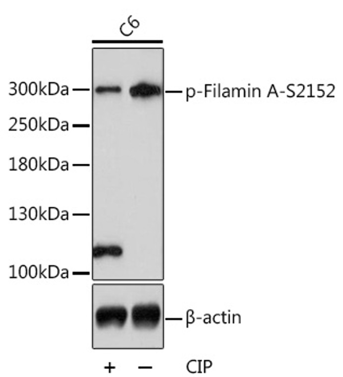
Rabbit polyclonal antibody anti-Phospho-FLNA-S2152 is suitable for use in Western Blot research applications. |
| Clonality: |
Polyclonal |
| Conjugation: |
Unconjugated |
| Isotype: |
IgG |
| Formulation: |
PBS with 0.01% Thimerosal, 50% Glycerol, pH7.3. |
| Purification: |
Affinity purification |
| Dilution Range: |
WB 1:500-1:2000 |
| Storage Instruction: |
Store at-20°C for up to 1 year from the date of receipt, and avoid repeat freeze-thaw cycles. |
| Gene Symbol: |
FLNA |
| Gene ID: |
2316 |
| Uniprot ID: |
FLNA_HUMAN |
| Immunogen: |
A synthetic phosphorylated peptide around S2152 of human Filamin A (NP_001104026.1). |
| Immunogen Sequence: |
APSVA |
| Tissue Specificity | Ubiquitous. |
| Post Translational Modifications | Phosphorylation at Ser-2152 is negatively regulated by the autoinhibited conformation of filamin repeats 19-21. Ligand binding induces a conformational switch triggering phosphorylation at Ser-2152 by PKA. Phosphorylation extent changes in response to cell activation. Polyubiquitination in the CH1 domain by a SCF-like complex containing ASB2 leads to proteasomal degradation. Prior dissociation from actin may be required to expose the target lysines. Ubiquitinated in endothelial cells by RNF213 downstream of the non-canonical Wnt signaling pathway, leading to its degradation by the proteasome. |
| Function | Promotes orthogonal branching of actin filaments and links actin filaments to membrane glycoproteins. Anchors various transmembrane proteins to the actin cytoskeleton and serves as a scaffold for a wide range of cytoplasmic signaling proteins. Interaction with FLNB may allow neuroblast migration from the ventricular zone into the cortical plate. Tethers cell surface-localized furin, modulates its rate of internalization and directs its intracellular trafficking. Involved in ciliogenesis. Plays a role in cell-cell contacts and adherens junctions during the development of blood vessels, heart and brain organs. Plays a role in platelets morphology through interaction with SYK that regulates ITAM- and ITAM-like-containing receptor signaling, resulting in by platelet cytoskeleton organization maintenance. During the axon guidance process, required for growth cone collapse induced by SEMA3A-mediated stimulation of neurons. |
| Protein Name | Filamin-AFln-AActin-Binding Protein 280Abp-280Alpha-FilaminEndothelial Actin-Binding ProteinFilamin-1Non-Muscle Filamin |
| Database Links | Reactome: R-HSA-114608Reactome: R-HSA-430116Reactome: R-HSA-446353Reactome: R-HSA-5627123Reactome: R-HSA-8983711 |
| Cellular Localisation | CytoplasmCell CortexCytoskeletonPerikaryonCell ProjectionGrowth ConeColocalizes With Cpmr1 In The Central Region Of Drg Neuron Growth ConeFollowing Sema3a Stimulation Of Drg NeuronsColocalizes With F-Actin |
| Alternative Antibody Names | Anti-Filamin-A antibodyAnti-Fln-A antibodyAnti-Actin-Binding Protein 280 antibodyAnti-Abp-280 antibodyAnti-Alpha-Filamin antibodyAnti-Endothelial Actin-Binding Protein antibodyAnti-Filamin-1 antibodyAnti-Non-Muscle Filamin antibodyAnti-FLNA antibodyAnti-FLN antibodyAnti-FLN1 antibody |
Information sourced from Uniprot.org
12 months for antibodies. 6 months for ELISA Kits. Please see website T&Cs for further guidance


