| Host: | Rabbit |
| Applications: | WB |
| Reactivity: | Human/Mouse/Rat |
| Note: | STRICTLY FOR FURTHER SCIENTIFIC RESEARCH USE ONLY (RUO). MUST NOT TO BE USED IN DIAGNOSTIC OR THERAPEUTIC APPLICATIONS. |
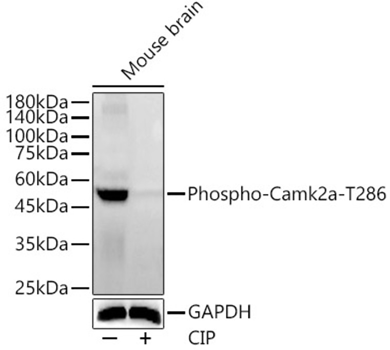
| Short Description: | Rabbit polyclonal antibody anti-Phospho-Camk2a-T286 is suitable for use in Western Blot research applications. |
| Clonality: | Polyclonal |
| Conjugation: | Unconjugated |
| Isotype: | IgG |
| Formulation: | PBS with 0.05% Proclin300, 50% Glycerol, pH7.3. |
| Purification: | Affinity purification |
| Dilution Range: | WB 1:2000-1:4000 |
| Storage Instruction: | Store at-20°C for up to 1 year from the date of receipt, and avoid repeat freeze-thaw cycles. |
| Gene Symbol: | CAMK2A |
| Gene ID: | 815 |
| Uniprot ID: | KCC2A_HUMAN |
| Immunogen: | A synthetic phosphorylated peptide around T286 of human Camk2a (NP_741960.1). |
| Immunogen Sequence: | QETVD |
| Post Translational Modifications | Autophosphorylation of Thr-286 following activation by Ca(2+)/calmodulin. Phosphorylation of Thr-286 locks the kinase into an activated state. Palmitoylated. Probably palmitoylated by ZDHHC3 and ZDHHC7. |
| Function | Calcium/calmodulin-dependent protein kinase that functions autonomously after Ca(2+)/calmodulin-binding and autophosphorylation, and is involved in various processes, such as synaptic plasticity, neurotransmitter release and long-term potentiation. Member of the NMDAR signaling complex in excitatory synapses, it regulates NMDAR-dependent potentiation of the AMPAR and therefore excitatory synaptic transmission. Regulates dendritic spine development. Also regulates the migration of developing neurons. Phosphorylates the transcription factor FOXO3 to activate its transcriptional activity. Phosphorylates the transcription factor ETS1 in response to calcium signaling, thereby decreasing ETS1 affinity for DNA. In response to interferon-gamma (IFN-gamma) stimulation, catalyzes phosphorylation of STAT1, stimulating the JAK-STAT signaling pathway. In response to interferon-beta (IFN-beta) stimulation, stimulates the JAK-STAT signaling pathway. Acts as a negative regulator of 2-arachidonoylglycerol (2-AG)-mediated synaptic signaling via modulation of DAGLA activity. |
| Protein Name | Calcium/Calmodulin-Dependent Protein Kinase Type Ii Subunit AlphaCam Kinase Ii Subunit AlphaCamk-Ii Subunit Alpha |
| Database Links | Reactome: R-HSA-111932Reactome: R-HSA-3371571Reactome: R-HSA-399719Reactome: R-HSA-4086398Reactome: R-HSA-438066Reactome: R-HSA-442982Reactome: R-HSA-5576892Reactome: R-HSA-5578775Reactome: R-HSA-5673000Reactome: R-HSA-5673001Reactome: R-HSA-6802946Reactome: R-HSA-6802952Reactome: R-HSA-6802955Reactome: R-HSA-877300Reactome: R-HSA-9022692Reactome: R-HSA-936837Reactome: R-HSA-9609736Reactome: R-HSA-9617324Reactome: R-HSA-9620244Reactome: R-HSA-9649948Reactome: R-HSA-9656223 |
| Cellular Localisation | SynapsePostsynaptic DensityCell ProjectionDendritic SpineDendritePostsynaptic Lipid Rafts |
| Alternative Antibody Names | Anti-Calcium/Calmodulin-Dependent Protein Kinase Type Ii Subunit Alpha antibodyAnti-Cam Kinase Ii Subunit Alpha antibodyAnti-Camk-Ii Subunit Alpha antibodyAnti-CAMK2A antibodyAnti-CAMKA antibodyAnti-KIAA0968 antibody |
Information sourced from Uniprot.org
12 months for antibodies. 6 months for ELISA Kits. Please see website T&Cs for further guidance


