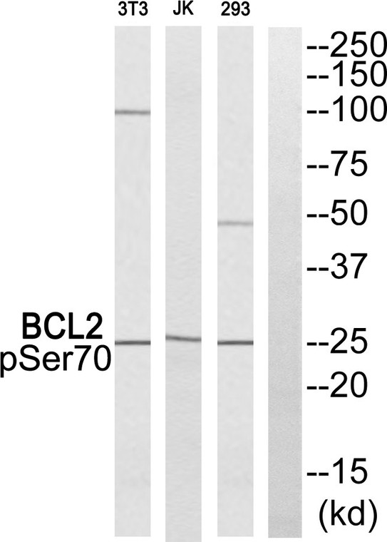
| Host: |
Rabbit |
| Applications: |
WB/ELISA |
| Reactivity: |
Mouse/Rat |
| Note: |
STRICTLY FOR FURTHER SCIENTIFIC RESEARCH USE ONLY (RUO). MUST NOT TO BE USED IN DIAGNOSTIC OR THERAPEUTIC APPLICATIONS. |
| Short Description: |
Rabbit polyclonal antibody anti-Phospho-BCL2-Ser70 (36-85 aa) is suitable for use in Western Blot and ELISA research applications. |
| Clonality: |
Polyclonal |
| Conjugation: |
Unconjugated |
| Isotype: |
IgG |
| Formulation: |
Liquid in PBS containing 50% Glycerol, 0.5% BSA and 0.02% Sodium Azide. |
| Purification: |
The antibody was affinity-purified from rabbit antiserum by affinity-chromatography using epitope-specific immunogen. |
| Concentration: |
1 mg/mL |
| Dilution Range: |
WB 1:500-1:2000ELISA 1:40000 |
| Storage Instruction: |
Store at-20°C for up to 1 year from the date of receipt, and avoid repeat freeze-thaw cycles. |
| Immunogen Region: |
36-85 aa |
| Specificity: |
Phospho-Bcl-2 (S70) Polyclonal Antibody detects endogenous levels of Bcl-2 protein only when phosphorylated at S70. |
| Immunogen: |
The antiserum was produced against synthesized peptide derived from the human BCL2 around the phosphorylation site of Ser70 at the amino acid range 36-85 |
| Post Translational Modifications | Phosphorylation/dephosphorylation on Ser-70 regulates anti-apoptotic activity. Growth factor-stimulated phosphorylation on Ser-70 by PKC is required for the anti-apoptosis activity and occurs during the G2/M phase of the cell cycle. In the absence of growth factors, BCL2 appears to be phosphorylated by other protein kinases such as ERKs and stress-activated kinases. Phosphorylated by MAPK8/JNK1 at Thr-69, Ser-70 and Ser-87, wich stimulates starvation-induced autophagy. Dephosphorylated by protein phosphatase 2A (PP2A). Proteolytically cleaved by caspases during apoptosis. The cleaved protein, lacking the BH4 motif, has pro-apoptotic activity, causes the release of cytochrome c into the cytosol promoting further caspase activity. Monoubiquitinated by PRKN, leading to increase its stability. Ubiquitinated by SCF(FBXO10), leading to its degradation by the proteasome. |
| Function | Suppresses apoptosis in a variety of cell systems including factor-dependent lymphohematopoietic and neural cells. Regulates cell death by controlling the mitochondrial membrane permeability. Appears to function in a feedback loop system with caspases. Inhibits caspase activity either by preventing the release of cytochrome c from the mitochondria and/or by binding to the apoptosis-activating factor (APAF-1). May attenuate inflammation by impairing NLRP1-inflammasome activation, hence CASP1 activation and IL1B release. |
| Protein Name | Apoptosis Regulator Bcl-2 |
| Database Links | Reactome: R-HSA-111447Reactome: R-HSA-111453Reactome: R-HSA-6785807Reactome: R-HSA-844455Reactome: R-HSA-9018519Reactome: R-HSA-9634638 |
| Cellular Localisation | Mitochondrion Outer MembraneSingle-Pass Membrane ProteinNucleus MembraneEndoplasmic Reticulum Membrane |
| Alternative Antibody Names | Anti-Apoptosis Regulator Bcl-2 antibodyAnti-BCL2 antibody |
Information sourced from Uniprot.org
12 months for antibodies. 6 months for ELISA Kits. Please see website T&Cs for further guidance


