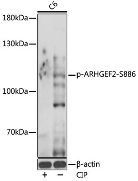
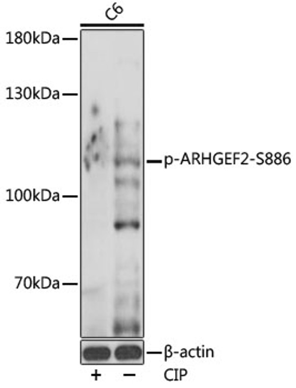
| Host: |
Rabbit |
| Applications: |
WB |
| Reactivity: |
Human/Mouse/Rat |
| Note: |
STRICTLY FOR FURTHER SCIENTIFIC RESEARCH USE ONLY (RUO). MUST NOT TO BE USED IN DIAGNOSTIC OR THERAPEUTIC APPLICATIONS. |
| Short Description: |
Rabbit polyclonal antibody anti-Phospho-ARHGEF2-S886 is suitable for use in Western Blot research applications. |
| Clonality: |
Polyclonal |
| Conjugation: |
Unconjugated |
| Isotype: |
IgG |
| Formulation: |
PBS with 0.01% Thimerosal, 50% Glycerol, pH7.3. |
| Purification: |
Affinity purification |
| Dilution Range: |
WB 1:500-1:2000 |
| Storage Instruction: |
Store at-20°C for up to 1 year from the date of receipt, and avoid repeat freeze-thaw cycles. |
| Gene Symbol: |
ARHGEF2 |
| Gene ID: |
9181 |
| Uniprot ID: |
ARHG2_HUMAN |
| Immunogen: |
A synthetic phosphorylated peptide around S886 of human ARHGEF2 (NP_001155855.1?). |
| Immunogen Sequence: |
RRSLP |
| Post Translational Modifications | Phosphorylation of Ser-886 by PAK1 induces binding to protein YWHAZ, promoting its relocation to microtubules and the inhibition of its activity. Phosphorylated by AURKA and CDK1 during mitosis, which negatively regulates its activity. Phosphorylation by MAPK1 or MAPK3 increases nucleotide exchange activity. Phosphorylation by PAK4 releases GEF-H1 from the microtubules. Phosphorylated on serine, threonine and tyrosine residues in a RIPK2-dependent manner. |
| Function | Activates Rho-GTPases by promoting the exchange of GDP for GTP. May be involved in epithelial barrier permeability, cell motility and polarization, dendritic spine morphology, antigen presentation, leukemic cell differentiation, cell cycle regulation, innate immune response, and cancer. Binds Rac-GTPases, but does not seem to promote nucleotide exchange activity toward Rac-GTPases, which was uniquely reported in PubMed:9857026. May stimulate instead the cortical activity of Rac. Inactive toward CDC42, TC10, or Ras-GTPases. Forms an intracellular sensing system along with NOD1 for the detection of microbial effectors during cell invasion by pathogens. Required for RHOA and RIP2 dependent NF-kappaB signaling pathways activation upon S.flexneri cell invasion. Involved not only in sensing peptidoglycan (PGN)-derived muropeptides through NOD1 that is independent of its GEF activity, but also in the activation of NF-kappaB by Shigella effector proteins (IpgB2 and OspB) which requires its GEF activity and the activation of RhoA. Involved in innate immune signaling transduction pathway promoting cytokine IL6/interleukin-6 and TNF-alpha secretion in macrophage upon stimulation by bacterial peptidoglycans.acts as a signaling intermediate between NOD2 receptor and RIPK2 kinase. Contributes to the tyrosine phosphorylation of RIPK2 through Src tyrosine kinase leading to NF-kappaB activation by NOD2. Overexpression activates Rho-, but not Rac-GTPases, and increases paracellular permeability. Involved in neuronal progenitor cell division and differentiation. Involved in the migration of precerebellar neurons. |
| Protein Name | Rho Guanine Nucleotide Exchange Factor 2Guanine Nucleotide Exchange Factor H1Gef-H1Microtubule-Regulated Rho-GefProliferating Cell Nucleolar Antigen P40 |
| Database Links | Reactome: R-HSA-193648Reactome: R-HSA-416482Reactome: R-HSA-8980692Reactome: R-HSA-9013026 |
| Cellular Localisation | CytoplasmCytoskeletonCell JunctionTight JunctionGolgi ApparatusSpindleCell ProjectionRuffle MembraneCytoplasmic VesicleLocalizes To The Tips Of Cortical Microtubules Of The Mitotic Spindle During Cell DivisionAnd Is Further Released Upon Microtubule DepolymerizationRecruited Into Membrane Ruffles Induced By SFlexneri At Tight Junctions Of Polarized Epithelial CellsColocalized With Nod2 And Ripk2 In Vesicles And With The Cytoskeleton |
| Alternative Antibody Names | Anti-Rho Guanine Nucleotide Exchange Factor 2 antibodyAnti-Guanine Nucleotide Exchange Factor H1 antibodyAnti-Gef-H1 antibodyAnti-Microtubule-Regulated Rho-Gef antibodyAnti-Proliferating Cell Nucleolar Antigen P40 antibodyAnti-ARHGEF2 antibodyAnti-KIAA0651 antibodyAnti-LFP40 antibody |
Information sourced from Uniprot.org
12 months for antibodies. 6 months for ELISA Kits. Please see website T&Cs for further guidance


