| Host: |
Rabbit |
| Applications: |
WB/IHC/IF |
| Reactivity: |
Human/Mouse/Rat |
| Note: |
STRICTLY FOR FURTHER SCIENTIFIC RESEARCH USE ONLY (RUO). MUST NOT TO BE USED IN DIAGNOSTIC OR THERAPEUTIC APPLICATIONS. |
| Short Description: |
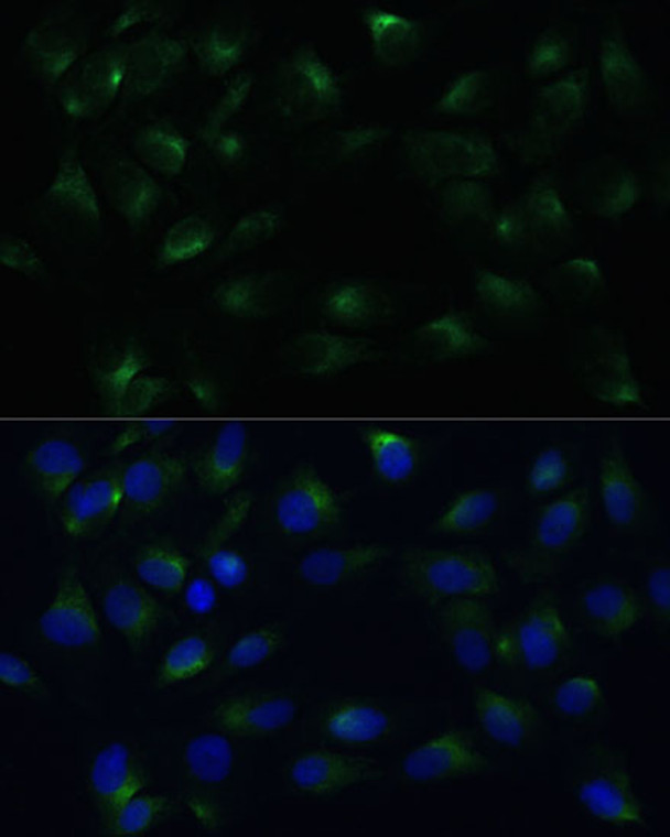
Rabbit polyclonal antibody anti-PGRMC1 (44-195) is suitable for use in Western Blot, Immunohistochemistry and Immunofluorescence research applications. |
| Clonality: |
Polyclonal |
| Conjugation: |
Unconjugated |
| Isotype: |
IgG |
| Formulation: |
PBS with 0.02% Sodium Azide, 50% Glycerol, pH7.3. |
| Purification: |
Affinity purification |
| Dilution Range: |
WB 1:500-1:2000IHC-P 1:50-1:100IF/ICC 1:50-1:100 |
| Storage Instruction: |
Store at-20°C for up to 1 year from the date of receipt, and avoid repeat freeze-thaw cycles. |
| Gene Symbol: |
PGRMC1 |
| Gene ID: |
10857 |
| Uniprot ID: |
PGRC1_HUMAN |
| Immunogen Region: |
44-195 |
| Immunogen: |
Recombinant fusion protein containing a sequence corresponding to amino acids 44-195 of human PGRMC1 (NP_006658.1). |
| Immunogen Sequence: |
KIVRGDQPAASGDSDDDEPP PLPRLKRRDFTPAELRRFDG VQDPRILMAINGKVFDVTKG RKFYGPEGPYGVFAGRDASR GLATFCLDKEALKDEYDDLS DLTAAQQETLSDWESQFTFK YHHVGKLLKEGEEPTVYSDE EEPKDESARKND |
| Tissue Specificity | Detected in urine (at protein level). Expressed by endometrial glands and stroma (at protein level). Widely expressed, with highest expression in liver and kidney. |
| Post Translational Modifications | O-glycosylated.contains chondroitin sulfate attached to Ser-54. Ser-54 is in the cytoplasmic domain but the glycosylated form was detected in urine, suggesting that the membrane-bound form is cleaved, allowing for production of a secreted form which is glycosylated. |
| Function | Component of a progesterone-binding protein complex. Binds progesterone. Has many reported cellular functions (heme homeostasis, interaction with CYPs). Required for the maintenance of uterine histoarchitecture and normal female reproductive lifespan. Intracellular heme chaperone. Regulates heme synthesis via interactions with FECH and acts as a heme donor for at least some hemoproteins. |
| Protein Name | Membrane-Associated Progesterone Receptor Component 1MprDap1Iza |
| Database Links | Reactome: R-HSA-6798695 |
| Cellular Localisation | Microsome MembraneSingle-Pass Membrane ProteinSmooth Endoplasmic Reticulum MembraneMitochondrion Outer MembraneExtracellular SideSecreted |
| Alternative Antibody Names | Anti-Membrane-Associated Progesterone Receptor Component 1 antibodyAnti-Mpr antibodyAnti-Dap1 antibodyAnti-Iza antibodyAnti-PGRMC1 antibodyAnti-HPR6.6 antibodyAnti-PGRMC antibody |
Information sourced from Uniprot.org
12 months for antibodies. 6 months for ELISA Kits. Please see website T&Cs for further guidance


