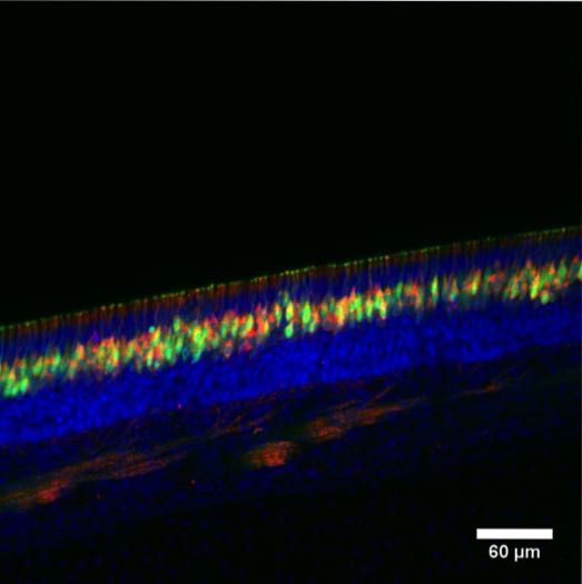
. Green: Olfactory sensory neurons. Red: Antibody immunoreactivity. Blue: Nuclei. PDE4A antibody labels the olfactory sensory neurons in mouse nasal epithelia.")
. Green: Olfactory sensory neurons. Red: Antibody immunoreactivity. Blue: Nuclei. PDE4A antibody labels the olfactory sensory neurons in mouse nasal epithelia.")
Anti-PDE4A antibody (C-Term) (STJ502279)
SPECIFICATIONS
ClonalityPolyclonal
HostRabbit
ConjugationUnconjugated
IsotypeIgG
ImmunogenSynthetic cyclic peptide corresponding to the C-terminal region of PDE4A. Common to all PDE4A proteins.
General Information
| Short Description | Rabbit polyclonal antibody anti-PDE4A (C-Term) is suitable for use in Confocal Microscopy, ELISA, Immunocytochemistry, Immunofluorescence, Immunohistochemistry, Immunoprecipitation and Western Blot research applications. |
| Applications | CM/ELISA/ICC/IF/IHC/IP/WB |
| Host | Rabbit |
| Reactivity | Human/Moose/Mouse/Rat |
| Note | STRICTLY FOR FURTHER SCIENTIFIC RESEARCH USE ONLY (RUO). MUST NOT TO BE USED IN DIAGNOSTIC OR THERAPEUTIC APPLICATIONS. |
Product Properties
| Clonality | Polyclonal |
| Isotype | IgG |
| Conjugation | Unconjugated |
| Concentration | 0.5 µg/µl |
| Purification | Affinity Purified |
| Dilution Range | WB: 1:1, 000-1:2, 500DB: 1:10, 000ELISA: 1:10, 000IP: 1:250IHC: 1:100-1:400ICC: 1:100-1:400IF: 1:100-1:400CFM: 1:100-1:400 |
| Formulation | Contains Tris, HCl/Glycine buffer pH 7.4-7.8, 30% Glycerol and 0.5% BSA, along with cryo-protective agents, Hepes, and long-term preservatives (0.02% Sodium Azide). |
| Storage Instruction | Store at-20°C for long term storage. Avoid freeze-thaw cycles. |
Target Information
| Gene Symbol | PDE4A |
| Gene ID | 5141 |
| Uniprot ID | PDE4A_HUMAN |
| Immunogen | Synthetic cyclic peptide corresponding to the C-terminal region of PDE4A. Common to all PDE4A proteins. |
| Immunogen Region | C-Term |
| Specificity | This antibody labels all known PDE4A variants including PDE4A1 (66 kDa) , PDE4A5 (109 kDa) , PDE4A8 (106 kDa) , AND PDE4Ax (10 kDa) and a 76 kDa testis specific A variant. |
Additional Info
| Tissue Specificity | Isoform 1: Expressed in lymphoid cell subsets including CD8-positive T cells and T-helper 2 cells. Expressed in dendritic cells. Isoform 2: Highly expressed in liver, stomach, testis, thyroid and adrenal glands and at a lower extent in placenta, kidney, pancreas, ovary, uterus and skin. Expressed in myeloid cell subsets including dendritic cells, monocytes, macrophages, eosinophils and mast cells. Expressed in natural killer cells. Expressed in bronchial smooth muscle. Isoform 6: Expressed at high levels in the heart and small intestine. It is also found in the brain, kidney, spleen, colon, salivary gland, ovary and peripheral blood lymphocytes. Isoform 7: Expressed predominantly in skeletal muscle and brain and at lower levels in the testis. Found in specific neuronal subpopulations including cortical pyramidal neurons, horn neurons in the spinal cord and Purkinje cells in cerebellum (at protein level). |
| Post Translational Modifications | Isoform 1: Proteolytically cleaved by CASP3. Isoform 2: Phosphorylated at Ser-119 by PKA. |
| Function | Hydrolyzes the second messenger 3',5'-cyclic AMP (cAMP), which is a key regulator of many important physiological processes. Isoform 1: Efficiently hydrolyzes cAMP. Isoform 2: Efficiently hydrolyzes cAMP. Isoform 3: Efficiently hydrolyzes cAMP. The phosphodiesterase activity is not affected by calcium, calmodulin or cyclic GMP (cGMP) levels. Does not hydrolyze cGMP. Isoform 4: Efficiently hydrolyzes cAMP. Isoform 6: Efficiently hydrolyzes cAMP. Isoform 7: Efficiently hydrolyzes cAMP. |
| Protein Name | 3' -5'-Cyclic-Amp Phosphodiesterase 4aDpde2Pde46Camp-Specific Phosphodiesterase 4a |
| Database Links | Reactome: R-HSA-180024Reactome: R-HSA-418555 |
| Cellular Localisation | Isoform 1: CytoplasmPerinuclear RegionIsoform 2: CytoplasmCell ProjectionRuffle MembraneIsoform 3: CytoplasmCytosolIsoform 4: MembranePeripheral Membrane ProteinIsoform 4 Has Propensity For Association With MembranesIsoform 6: CytoplasmIsoform 7: CytoplasmMembranePredominantly Cytosolic |
| Alternative Antibody Names | Anti-3' -5'-Cyclic-Amp Phosphodiesterase 4a antibodyAnti-Dpde2 antibodyAnti-Pde46 antibodyAnti-Camp-Specific Phosphodiesterase 4a antibodyAnti-PDE4A antibodyAnti-DPDE2 antibody |
Information sourced from Uniprot.org