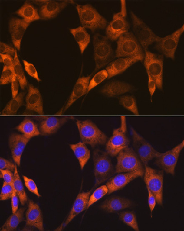
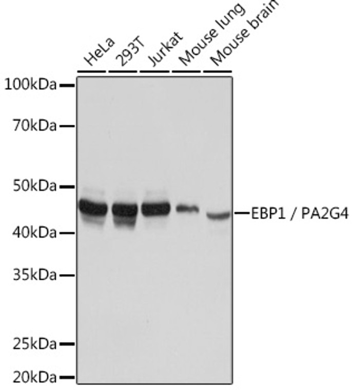
| Host: |
Rabbit |
| Applications: |
WB/IHC/IF |
| Reactivity: |
Human/Mouse/Rat |
| Note: |
STRICTLY FOR FURTHER SCIENTIFIC RESEARCH USE ONLY (RUO). MUST NOT TO BE USED IN DIAGNOSTIC OR THERAPEUTIC APPLICATIONS. |
| Short Description: |
Rabbit monoclonal antibody anti-EBP1/PA2G4 (300-394) is suitable for use in Western Blot, Immunohistochemistry and Immunofluorescence research applications. |
| Clonality: |
Monoclonal |
| Clone ID: |
S9MR |
| Conjugation: |
Unconjugated |
| Isotype: |
IgG |
| Formulation: |
PBS with 0.02% Sodium Azide, 0.05% BSA, 50% Glycerol, pH7.3. |
| Purification: |
Affinity purification |
| Dilution Range: |
WB 1:500-1:1000IHC-P 1:50-1:200IF/ICC 1:50-1:200 |
| Storage Instruction: |
Store at-20°C for up to 1 year from the date of receipt, and avoid repeat freeze-thaw cycles. |
| Gene Symbol: |
PA2G4 |
| Gene ID: |
5036 |
| Uniprot ID: |
PA2G4_HUMAN |
| Immunogen Region: |
300-394 |
| Immunogen: |
Recombinant fusion protein containing a sequence corresponding to amino acids 300-394 of human EBP1/PA2G4 (NP_006182.2). |
| Immunogen Sequence: |
ELLQPFNVLYEKEGEFVAQF KFTVLLMPNGPMRITSGPFE PDLYKSEMEVQDAELKALLQ SSASRKTQKKKKKKASKTAE NATSGETLEENEAGD |
| Tissue Specificity | Isoform 2 is undetectable whereas isoform 1 is strongly expressed in cancer cells (at protein level). Isoform 1 and isoform 2 are widely expressed, including heart, brain, lung, pancreas, skeletal muscle, kidney, placenta and liver. |
| Post Translational Modifications | Phosphorylated on serine and threonine residues. Phosphorylation is enhanced by HRG treatment. Basal phosphorylation is PKC-dependent and HRG-induced phosphorylation is predominantly PKC-independent. Phosphorylation at Ser-361 by PKC/PRKCD regulates its nucleolar localization. In cancer cells, isoform 2 is polyubiquitinated leading to its proteasomal degradation and phosphorylation by PKC/PRKCD enhances polyubiquitination. |
| Function | May play a role in a ERBB3-regulated signal transduction pathway. Seems be involved in growth regulation. Acts a corepressor of the androgen receptor (AR) and is regulated by the ERBB3 ligand neuregulin-1/heregulin (HRG). Inhibits transcription of some E2F1-regulated promoters, probably by recruiting histone acetylase (HAT) activity. Binds RNA. Associates with 28S, 18S and 5.8S mature rRNAs, several rRNA precursors and probably U3 small nucleolar RNA. May be involved in regulation of intermediate and late steps of rRNA processing. May be involved in ribosome assembly. Mediates cap-independent translation of specific viral IRESs (internal ribosomal entry site). Regulates cell proliferation, differentiation, and survival. Isoform 1 suppresses apoptosis whereas isoform 2 promotes cell differentiation. |
| Protein Name | Proliferation-Associated Protein 2g4Cell Cycle Protein P38-2g4 HomologHg4-1Erbb3-Binding Protein 1 |
| Database Links | Reactome: R-HSA-6798695 |
| Cellular Localisation | Isoform 1: CytoplasmNucleusNucleolusTranslocates To The Nucleus Upon Treatment With HrgPhosphorylation At Ser-361 By Pkc/Prkcd Regulates Its Nucleolar LocalizationIsoform 2: Cytoplasm |
| Alternative Antibody Names | Anti-Proliferation-Associated Protein 2g4 antibodyAnti-Cell Cycle Protein P38-2g4 Homolog antibodyAnti-Hg4-1 antibodyAnti-Erbb3-Binding Protein 1 antibodyAnti-PA2G4 antibodyAnti-EBP1 antibody |
Information sourced from Uniprot.org
12 months for antibodies. 6 months for ELISA Kits. Please see website T&Cs for further guidance


![Western blot analysis of lysates from wild type (WT) and EBP1/PA2G4 knockout (KO) 293T cells, using [KO Validated] EBP1/PA2G4 Rabbit polyclonal antibody (STJ11100848) at 1:1000 dilution. Secondary antibody: HRP Goat Anti-Rabbit IgG (H+L) (STJS000856) at 1:10000 dilution. Lysates/proteins: 25 Mu g per lane. Blocking buffer: 3% nonfat dry milk in TBST. Detection: ECL Basic Kit. Exposure time: 5s. Western blot analysis of lysates from wild type (WT) and EBP1/PA2G4 knockout (KO) 293T cells, using [KO Validated] EBP1/PA2G4 Rabbit polyclonal antibody (STJ11100848) at 1:1000 dilution. Secondary antibody: HRP Goat Anti-Rabbit IgG (H+L) (STJS000856) at 1:10000 dilution. Lysates/proteins: 25 Mu g per lane. Blocking buffer: 3% nonfat dry milk in TBST. Detection: ECL Basic Kit. Exposure time: 5s.](https://cdn11.bigcommerce.com/s-zso2xnchw9/images/stencil/300x300/products/89782/358253/STJ11100848_1__80011.1713122590.jpg?c=1)

