 staining of Rat Pancreas lysate (35µg protein in RIPA buffer). Detected by chemiluminescence.")
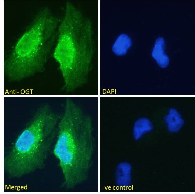
 followed by Alexa Fluor 488 secondary antibody (2ug/ml) , showing nuclear and cytoplasmic staining. The nuclear stain is DAPI (blue). Negative control: Unimmunized goat IgG (10ug/ml) followed by Alexa Fluor 488 secondary antibody (2ug/ml).")
 followed by Alexa Fluor 488 secondary antibody (2ug/ml) , showing nuclear staining. Actin filaments were stained with phalloidin (red) and the nuclear stain is DAPI (blue). Negative control: Unimmunized goat IgG (10ug/ml) followed by Alexa Fluor 488 secondary antibody (2ug/ml).")
 followed by Alexa Fluor 488 secondary antibody (2ug/ml) , showing nuclear and membrane/cytoplasmic staining. Actin filaments were stained with phalloidin (red) and the nuclear stain is DAPI (blue). Negative control: Unimmunized goat IgG (10ug/ml) followed by Alexa Fluor 488 secondary antibody (2ug/ml).")
 , permeabilized with 0. 5% Triton. Primary incubation 1hr (10ug/ml) followed by Alexa Fluor 488 secondary antibody (1ug/ml). IgG control: Unimmunized goat IgG (black line) followed by Alexa Fluor 488 secondary antibody.")
 staining of Rat Pancreas lysate (35µg protein in RIPA buffer). Detected by chemiluminescence.")
Anti-OGT antibody (Internal) (STJ71101)
SPECIFICATIONS
ClonalityPolyclonal
HostGoat
ConjugationUnconjugated
IsotypeIgG
General Information
| Short Description | Goat polyclonal antibody anti-OGT (Internal) is suitable for use in ELISA, Western Blot, Immunohistochemistry, Immunofluorescence and Flow Cytometry research applications. |
| Applications | Pep-ELISA/WB/IHC/IF/FC |
| Host | Goat |
| Reactivity | Human/Mouse/Rat/Dog/Cow |
| Note | STRICTLY FOR FURTHER SCIENTIFIC RESEARCH USE ONLY (RUO). MUST NOT TO BE USED IN DIAGNOSTIC OR THERAPEUTIC APPLICATIONS. |
Product Properties
| Clonality | Polyclonal |
| Isotype | IgG |
| Conjugation | Unconjugated |
| Concentration | 0.5 mg/mL |
| Purification | Purified from goat serum by ammonium sulphate precipitation followed by antigen affinity chromatography using the immunizing peptide. |
| Dilution Range | Peptide ELISA: antibody detection limit dilution 1:64000.WB: Approx 110kDa band observed in Rat Pancreas lysates calculated MW of 116kDa according to NP_858058.2). An additional band of unknown identity was also consistently observed at 60kDa. T |
| Formulation | 0.5 mg/ml in Tris saline, 0.02% sodium azide, pH7.3 with 0.5% bovine serum albumin. NA |
| Storage Instruction | Store at-20°C on receipt and minimise freeze-thaw cycles. |
Target Information
| Gene Symbol | OGT |
| Gene ID | 8473 |
| Uniprot ID | OGT1_HUMAN |
| Accession Number | NP_858058.1; NP_858059.1 |
| Immunogen Region | Internal |
| Immunogen Sequence | YEHPKDLKLSDGR |
| Specificity | This antibody is expected to recognise both reported isoforms (NP_858058.1 and NP_858059.1 |
Additional Info
| Post Translational Modifications | Ubiquitinated, leading to its proteasomal degradation. Phosphorylation on Ser-3 or Ser-4 by GSK3-beta positively regulates its activity. Phosphorylation at Thr-454 by AMPK promotes nuclear localization. Glycosylated via autocatalysis.O-GlcNAcylation at Ser-399 promotes nuclear localization. Isoform 4: Glycosylated via autocatalysis.does not affect the enzyme activity but regulates substrate selectivity. |
| Function | Catalyzes the transfer of a single N-acetylglucosamine from UDP-GlcNAc to a serine or threonine residue in cytoplasmic and nuclear proteins resulting in their modification with a beta-linked N-acetylglucosamine (O-GlcNAc). Glycosylates a large and diverse number of proteins including histone H2B, AKT1, AMPK, ATG4B, CAPRIN1, EZH2, FNIP1, GSDMD, KRT7, LMNA, LMNB1, LMNB2, RPTOR, HOXA1, PFKL, KMT2E/MLL5, MAPT/TAU, TET2, RBL2, RET, NOD2 and HCFC1. Can regulate their cellular processes via cross-talk between glycosylation and phosphorylation or by affecting proteolytic processing. Involved in insulin resistance in muscle and adipocyte cells via glycosylating insulin signaling components and inhibiting the 'Thr-308' phosphorylation of AKT1, enhancing IRS1 phosphorylation and attenuating insulin signaling. Involved in glycolysis regulation by mediating glycosylation of 6-phosphofructokinase PFKL, inhibiting its activity. Plays a key role in chromatin structure by mediating O-GlcNAcylation of 'Ser-112' of histone H2B: recruited to CpG-rich transcription start sites of active genes via its interaction with TET proteins (TET1, TET2 or TET3). As part of the NSL complex indirectly involved in acetylation of nucleosomal histone H4 on several lysine residues. O-GlcNAcylation of 'Ser-75' of EZH2 increases its stability, and facilitating the formation of H3K27me3 by the PRC2/EED-EZH2 complex. Stabilizes KMT2E/MLL5 by mediating its glycosylation, thereby preventing KMT2E/MLL5 ubiquitination. Regulates circadian oscillation of the clock genes and glucose homeostasis in the liver. Stabilizes clock proteins BMAL1 and CLOCK through O-glycosylation, which prevents their ubiquitination and subsequent degradation. Promotes the CLOCK-BMAL1-mediated transcription of genes in the negative loop of the circadian clock such as PER1/2 and CRY1/2. O-glycosylates HCFC1 and regulates its proteolytic processing and transcriptional activity. Component of a THAP1/THAP3-HCFC1-OGT complex that is required for the regulation of the transcriptional activity of RRM1. Regulates mitochondrial motility in neurons by mediating glycosylation of TRAK1. Promotes autophagy by mediating O-glycosylation of ATG4B. Acts as a regulator of mTORC1 signaling by mediating O-glycosylation of RPTOR and FNIP1: O-GlcNAcylation of RPTOR in response to glucose sufficiency promotes activation of the mTORC1 complex. Isoform 2: The mitochondrial isoform (mOGT) is cytotoxic and triggers apoptosis in several cell types including INS1, an insulinoma cell line. Isoform 4: Has N-acetylglucosaminyltransferase activity: glycosylates proteins, such as HNRNPU, NEUROD1, NUP62 and PDCD6IP. Displays specific substrate selectivity compared to other isoforms. |
| Protein Name | Udp-N-Acetylglucosamine--Peptide N-Acetylglucosaminyltransferase 110 Kda SubunitO-Glcnac Transferase Subunit P110O-Linked N-Acetylglucosamine Transferase 110 Kda SubunitOgt |
| Database Links | Reactome: R-HSA-3214847Reactome: R-HSA-5213460Reactome: R-HSA-5675482Reactome: R-HSA-5689603Reactome: R-HSA-9772755 |
| Cellular Localisation | NucleusCytoplasmPredominantly Localizes To The NucleusTranslocates Into The Nucleus Via Association With Importin Kpna1Isoform 2: MitochondrionMembraneAssociates With The Mitochondrial Inner MembraneIsoform 3: CytoplasmCell MembraneMitochondrion MembraneCell ProjectionMostly In The NucleusRetained In The Nucleus Via Interaction With Hcfc1After Insulin InductionTranslocated From The Nucleus To The Cell Membrane Via Phosphatidylinositide BindingColocalizes With Akt1 At The Plasma MembraneTrak1 Recruits This Protein To MitochondriaIn The Absence Of Trak1Localizes In Cytosol And NucleusIsoform 4: Cytoplasm |
| Alternative Antibody Names | Anti-Udp-N-Acetylglucosamine--Peptide N-Acetylglucosaminyltransferase 110 Kda Subunit antibodyAnti-O-Glcnac Transferase Subunit P110 antibodyAnti-O-Linked N-Acetylglucosamine Transferase 110 Kda Subunit antibodyAnti-Ogt antibodyAnti-OGT antibody |
Information sourced from Uniprot.org