| Host: |
Rabbit |
| Applications: |
WB/IF/IP |
| Reactivity: |
Human/Mouse/Rat |
| Note: |
STRICTLY FOR FURTHER SCIENTIFIC RESEARCH USE ONLY (RUO). MUST NOT TO BE USED IN DIAGNOSTIC OR THERAPEUTIC APPLICATIONS. |
| Short Description: |
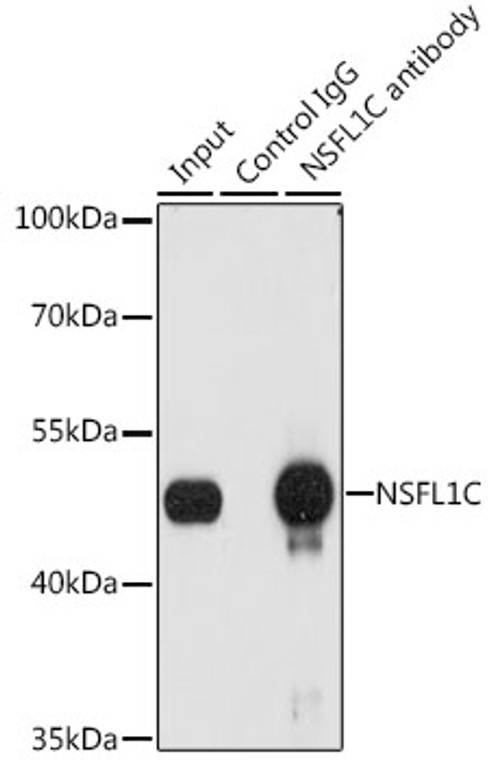
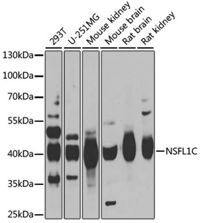
Rabbit polyclonal antibody anti-NSFL1C (1-185) is suitable for use in Western Blot, Immunofluorescence and Immunoprecipitation research applications. |
| Clonality: |
Polyclonal |
| Conjugation: |
Unconjugated |
| Isotype: |
IgG |
| Formulation: |
PBS with 0.02% Sodium Azide, 50% Glycerol, pH7.3. |
| Purification: |
Affinity purification |
| Dilution Range: |
WB 1:500-1:2000IF/ICC 1:50-1:200IP 1:50-1:100 |
| Storage Instruction: |
Store at-20°C for up to 1 year from the date of receipt, and avoid repeat freeze-thaw cycles. |
| Gene Symbol: |
NSFL1C |
| Gene ID: |
55968 |
| Uniprot ID: |
NSF1C_HUMAN |
| Immunogen Region: |
1-185 |
| Immunogen: |
Recombinant fusion protein containing a sequence corresponding to amino acids 1-185 of human NSFL1C (NP_057227.2). |
| Immunogen Sequence: |
MAAERQEALREFVAVTGAEE DRARFFLESAGWDLQIALAS FYEDGGDEDIVTISQATPSS VSRGTAPSDNRVTSFRDLIH DQDEDEEEEEGQRFYAGGSE RSGQQIVGPPRKKSPNELVD DLFKGAKEHGAVAVERVTKS PGETSKPRPFAGGGYRLGAA PEEESAYVAGEKRQHSSQDV HVVLK |
| Post Translational Modifications | Phosphorylated during mitosis. Phosphorylation inhibits interaction with Golgi membranes and is required for the fragmentation of the Golgi stacks during mitosis. |
| Function | Reduces the ATPase activity of VCP. Necessary for the fragmentation of Golgi stacks during mitosis and for VCP-mediated reassembly of Golgi stacks after mitosis. May play a role in VCP-mediated formation of transitional endoplasmic reticulum (tER). Inhibits the activity of CTSL (in vitro). Together with UBXN2B/p37, regulates the centrosomal levels of kinase AURKA/Aurora A during mitotic progression by promoting AURKA removal from centrosomes in prophase. Also, regulates spindle orientation during mitosis. |
| Protein Name | Nsfl1 Cofactor P47Ubx Domain-Containing Protein 2cP97 Cofactor P47 |
| Database Links | Reactome: R-HSA-9013407 |
| Cellular Localisation | NucleusGolgi ApparatusGolgi StackChromosomeCytoplasmCytoskeletonMicrotubule Organizing CenterCentrosomePredominantly Nuclear In Interphase CellsBound To The Axial Elements Of Sex Chromosomes In Pachytene SpermatocytesA Small Proportion Of The Protein Is CytoplasmicAssociated With Golgi StacksLocalizes To Centrosome During Mitotic Prophase And Metaphase |
| Alternative Antibody Names | Anti-Nsfl1 Cofactor P47 antibodyAnti-Ubx Domain-Containing Protein 2c antibodyAnti-P97 Cofactor P47 antibodyAnti-NSFL1C antibodyAnti-UBXN2C antibody |
Information sourced from Uniprot.org
12 months for antibodies. 6 months for ELISA Kits. Please see website T&Cs for further guidance


