| Host: |
Rabbit |
| Applications: |
WB |
| Reactivity: |
Human |
| Note: |
STRICTLY FOR FURTHER SCIENTIFIC RESEARCH USE ONLY (RUO). MUST NOT TO BE USED IN DIAGNOSTIC OR THERAPEUTIC APPLICATIONS. |
| Short Description: |
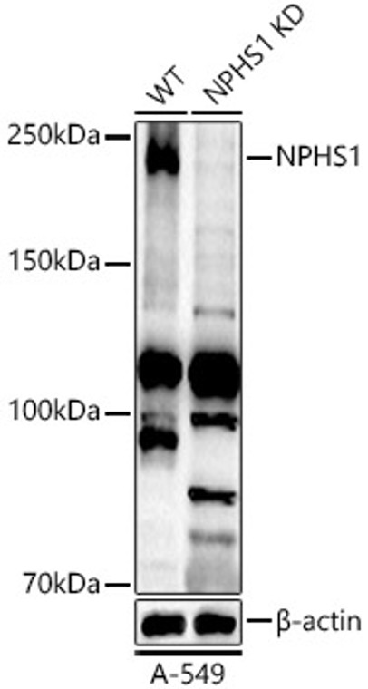
Rabbit polyclonal antibody anti-Nephrin (1019-1241) is suitable for use in Western Blot research applications. |
| Clonality: |
Polyclonal |
| Conjugation: |
Unconjugated |
| Isotype: |
IgG |
| Formulation: |
PBS with 0.05% Proclin300, 50% Glycerol, pH7.3. |
| Purification: |
Affinity purification |
| Dilution Range: |
WB 1:500-1:1000 |
| Storage Instruction: |
Store at-20°C for up to 1 year from the date of receipt, and avoid repeat freeze-thaw cycles. |
| Gene Symbol: |
NPHS1 |
| Gene ID: |
4868 |
| Uniprot ID: |
NPHN_HUMAN |
| Immunogen Region: |
1019-1241 |
| Immunogen: |
Recombinant fusion protein containing a sequence corresponding to amino acids 1019-1241 of human NPHS1 (NP_004637.1). |
| Immunogen Sequence: |
LGDSGLADKGTQLPITTPGL HQPSGEPEDQLPTEPPSGPS GLPLLPVLFALGGLLLLSNA SCVGGVLWQRRLRRLAEGIS EKTEAGSEEDRVRNEYEESQ WTGERDTQSSTVSTTEAEPY YRSLRDFSPQLPPTQEEVSY SRGFTGEDEDMAFPGHLYDE VERTYPPSGAWGPLYDEVQM GPWDLHWPEDTYQDPRGIYD QVAGDLDTLEPDSLPFELRG HLV |
| Tissue Specificity | Specifically expressed in podocytes of kidney glomeruli. |
| Post Translational Modifications | Phosphorylated at Tyr-1193 by FYN, leading to the recruitment and activation of phospholipase C-gamma-1/PLCG1. Tyrosine phosphorylation is reduced by high glucose levels. Dephosphorylated by tensin TNS2 which leads to reduced binding of NPHN1 to PIK3R1. |
| Function | Seems to play a role in the development or function of the kidney glomerular filtration barrier. Regulates glomerular vascular permeability. May anchor the podocyte slit diaphragm to the actin cytoskeleton. Plays a role in skeletal muscle formation through regulation of myoblast fusion. |
| Protein Name | NephrinRenal Glomerulus-Specific Cell Adhesion Receptor |
| Database Links | Reactome: R-HSA-373753 |
| Cellular Localisation | Cell MembraneSingle-Pass Type I Membrane ProteinPredominantly Located At Podocyte Slit Diaphragm Between Podocyte Foot ProcessesAlso Associated With Podocyte Apical Plasma Membrane |
| Alternative Antibody Names | Anti-Nephrin antibodyAnti-Renal Glomerulus-Specific Cell Adhesion Receptor antibodyAnti-NPHS1 antibodyAnti-NPHN antibody |
Information sourced from Uniprot.org
12 months for antibodies. 6 months for ELISA Kits. Please see website T&Cs for further guidance


