| Host: |
Rabbit |
| Applications: |
WB |
| Reactivity: |
Human/Mouse |
| Note: |
STRICTLY FOR FURTHER SCIENTIFIC RESEARCH USE ONLY (RUO). MUST NOT TO BE USED IN DIAGNOSTIC OR THERAPEUTIC APPLICATIONS. |
| Short Description: |
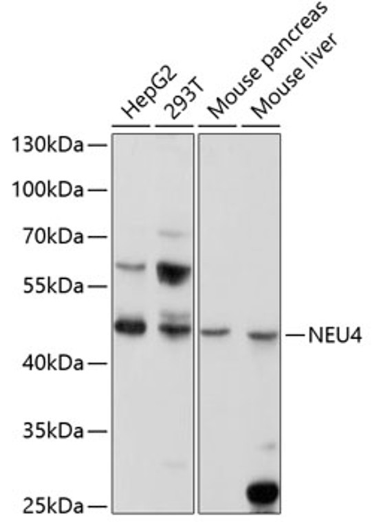
Rabbit polyclonal antibody anti-NEU4 (197-496) is suitable for use in Western Blot research applications. |
| Clonality: |
Polyclonal |
| Conjugation: |
Unconjugated |
| Isotype: |
IgG |
| Formulation: |
PBS with 0.02% Sodium Azide, 50% Glycerol, pH7.3. |
| Purification: |
Affinity purification |
| Dilution Range: |
WB 1:500-1:2000 |
| Storage Instruction: |
Store at-20°C for up to 1 year from the date of receipt, and avoid repeat freeze-thaw cycles. |
| Gene Symbol: |
NEU4 |
| Gene ID: |
129807 |
| Uniprot ID: |
NEUR4_HUMAN |
| Immunogen Region: |
197-496 |
| Immunogen: |
Recombinant fusion protein containing a sequence corresponding to amino acids 197-496 of human NEU4 (NP_542779.2). |
| Immunogen Sequence: |
ECFGKICRTSPHSFAFYSDD HGRTWRCGGLVPNLRSGECQ LAAVDGGQAGSFLYCNARSP LGSRVQALSTDEGTSFLPAE RVASLPETAWGCQGSIVGFP APAPNRPRDDSWSVGPGSPL QPPLLGPGVHEPPEEAAVDP RGGQVPGGPFSRLQPRGDGP RQPGPRPGVSGDVGSWTLAL PMPFAAPPQSPTWLLYSHPV GRRARLHMGIRLSQSPLDPR SWTEPWVIYEGPSGYSDLA |
| Tissue Specificity | Isoform 1: Predominant form in liver. Also expressed in brain, kidney and colon. Isoform 2: Highly expressed in brain and at lower levels in kidney and liver. |
| Post Translational Modifications | Isoform 2: N-glycosylated. |
| Function | Exo-alpha-sialidase that catalyzes the hydrolytic cleavage of the terminal sialic acid (N-acetylneuraminic acid, Neu5Ac) of a glycan moiety in the catabolism of glycolipids, glycoproteins and oligosacharides. Efficiently hydrolyzes gangliosides including alpha-(2->3)-sialylated GD1a and GM3 and alpha-(2->8)-sialylated GD3. Hydrolyzes poly-alpha-(2->8)-sialylated neural cell adhesion molecule NCAM1 likely at growth cones, suppressing neurite outgrowth in hippocampal neurons. May desialylate sialyl Lewis A and X antigens at the cell surface, down-regulating these glycan epitopes recognized by SELE/E selectin in the initiation of cell adhesion and extravasation. Has sialidase activity toward mucin, fetuin and sialyllactose. |
| Protein Name | Sialidase-4N-Acetyl-Alpha-Neuraminidase 4 |
| Database Links | Reactome: R-HSA-1660662 Q8WWR8-2Reactome: R-HSA-4085001 |
| Cellular Localisation | Isoform 1: Cell MembranePeripheral Membrane ProteinEndoplasmic Reticulum MembraneMicrosome MembraneMitochondrion MembraneCell ProjectionNeuron ProjectionPredominantly Associates With Endoplasmic Reticulum MembranesOnly A Small Fraction Associates With Mitochondrial And Plasma MembranesIsoform 2: Mitochondrion Inner MembraneMitochondrion Outer MembraneLysosome LumenIsoform 2 Is SolubleN-Glycosylated And Found In The Lumen Of LysosomesHoweverNo Signal Sequence Nor N-Glycosylation Site Is Predicted From The Sequence |
| Alternative Antibody Names | Anti-Sialidase-4 antibodyAnti-N-Acetyl-Alpha-Neuraminidase 4 antibodyAnti-NEU4 antibodyAnti-LP5125 antibody |
Information sourced from Uniprot.org
12 months for antibodies. 6 months for ELISA Kits. Please see website T&Cs for further guidance


