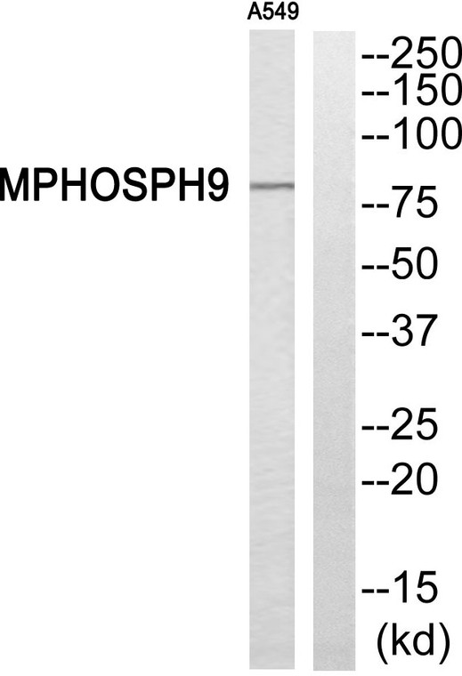
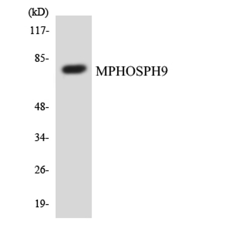
| Host: |
Rabbit |
| Applications: |
WB/IHC |
| Reactivity: |
Human/Rat/Mouse |
| Note: |
STRICTLY FOR FURTHER SCIENTIFIC RESEARCH USE ONLY (RUO). MUST NOT TO BE USED IN DIAGNOSTIC OR THERAPEUTIC APPLICATIONS. |
| Short Description: |
Rabbit polyclonal antibody anti-M-phase phosphoprotein 9 (539-588) is suitable for use in Western Blot and Immunohistochemistry research applications. |
| Clonality: |
Polyclonal |
| Conjugation: |
Unconjugated |
| Isotype: |
IgG |
| Formulation: |
Liquid in PBS containing 50% Glycerol, 0.5% BSA and 0.02% Sodium Azide. |
| Purification: |
The antibody was affinity-purified from rabbit antiserum by affinity-chromatography using epitope-specific immunogen. |
| Concentration: |
1 mg/mL |
| Dilution Range: |
WB 1:500-2000IHC-p 1:50-300 |
| Storage Instruction: |
Store at-20°C for up to 1 year from the date of receipt, and avoid repeat freeze-thaw cycles. |
| Gene Symbol: |
MPHOSPH9 |
| Gene ID: |
10198 |
| Uniprot ID: |
MPP9_HUMAN |
| Immunogen Region: |
539-588 |
| Specificity: |
MPP9 Polyclonal Antibody detects endogenous levels of MPP9 protein. |
| Immunogen: |
The antiserum was produced against synthesized peptide derived from human MPHOSPH9. AA range:539-588 |
| Post Translational Modifications | TTBK2-mediated phosphorylation at Ser-781 and Ser-788, promotes its ubiquitination at Lys-784 leading to proteasomal degradation, loss of MPHOSPH9 facilitates the removal of the CP110-CEP97 complex from the mother centrioles, promoting the initiation of ciliogenesis. Phosphorylated in M (mitotic) phase. Ubiquitinated at Lys-784, leading to proteasomal degradation. |
| Function | Negatively regulates cilia formation by recruiting the CP110-CEP97 complex (a negative regulator of ciliogenesis) at the distal end of the mother centriole in ciliary cells. At the beginning of cilia formation, MPHOSPH9 undergoes TTBK2-mediated phosphorylation and degradation via the ubiquitin-proteasome system and removes itself and the CP110-CEP97 complex from the distal end of the mother centriole, which subsequently promotes cilia formation. |
| Protein Name | M-Phase Phosphoprotein 9 |
| Cellular Localisation | CytoplasmCytoskeletonMicrotubule Organizing CenterCentrosomeCentrioleGolgi Apparatus MembranePeripheral Membrane ProteinLocalizes To The Distal And Proximal End Of Centriole Pairs In Duplicated CentrosomesIn Ciliated CellsLocalizes To The Distal And Proximal End Of Daughter Centriole And Proximal Of The Mother Centriole But Not In The Distal End Of The Mother CentrioleRecruited By Kif24 To The Distal End Of Mother Centriole Where It Forms A Ring-Like Structure |
| Alternative Antibody Names | Anti-M-Phase Phosphoprotein 9 antibodyAnti-MPHOSPH9 antibodyAnti-MPP9 antibody |
Information sourced from Uniprot.org
12 months for antibodies. 6 months for ELISA Kits. Please see website T&Cs for further guidance


