• CUT&Tag was performed using the CUT&Tag Assay Kit (pAG-Tn5) for Illumina (RK20265) from 10⁵ K562 cells with 1 Mu g MonoMethyl-Histone H3-K9 antibody (STJ11103432) , along with a Goat Anti-rabbit IgG (H+L). The CUT&Tag results indicate the enrichment pattern of H3K9me1 in representative gene loci (MYOD1) , as shown in figure.
  • Western blot analysis of extracts from various cell lines, using MonoMethyl-Histone H3-K9 antibody (STJ11103432) at 1:1000 dilution. Secondary antibody: HRP Goat Anti-rabbit IgG (H+L) (STJS000856) at 1:10000 dilution. Lysates/proteins: 25 Mu g per lane. Blocking buffer: 3% non-fat dry milk in TBST. Detection: ECL Basic Kit. Exposure time: 10s.
  • Dot-blot analysis of all sorts of peptides using MonoMethyl-Histone H3-K9 antibody (STJ11103432) at 1:1000 dilution.
  • Immunohistochemistry analysis of paraffin-embedded human colon carcinoma using MonoMethyl-Histone H3-K9 antibody (STJ11103432) at dilution of 1:100 (40x lens). Perform high pressure antigen retrieval with 10 mM citrate buffer pH 6. 0 before commencing with immunohistochemistry staining protocol.
  • Immunohistochemistry analysis of paraffin-embedded mouse lung using MonoMethyl-Histone H3-K9 antibody (STJ11103432) at dilution of 1:100 (40x lens). Perform high pressure antigen retrieval with 10 mM citrate buffer pH 6. 0 before commencing with immunohistochemistry staining protocol.
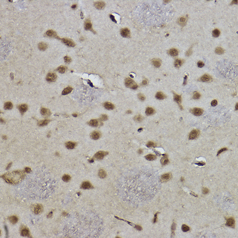
  • Immunohistochemistry analysis of paraffin-embedded rat brain using MonoMethyl-Histone H3-K9 antibody (STJ11103432) at dilution of 1:100 (40x lens). Perform high pressure antigen retrieval with 10 mM citrate buffer pH 6. 0 before commencing with immunohistochemistry staining protocol.
  • Confocal imaging of U-2 OS cells using MonoMethyl-Histone H3-K9 rabbit monoclonal antibody (STJ11103432, dilution 1:100) (Red). The cells were counterstained with Alpha-Tubulin mouse monoclonal antibody (dilution 1:400) (Green). DAPI was used for nuclear staining (blue). Objective: 100x.
  • Chromatin immunoprecipitation analysis of extracts from HeLa cells, using MonoMethyl-Histone H3-K9 antibody (STJ11103432) and rabbit IgG. The amount of immunoprecipitated DNA was checked by quantitative PCR. Histogram was constructed by the ratios of the immunoprecipitated DNA to the input.

Anti-Mono-Methyl-Histone H3-K9 antibody [S2MR] (STJ11103432)

SKU:
STJ11103432

Shipping:
Free Shipping
Current Stock:
Host: Rabbit
Applications: WB/IHC/IF
Reactivity: Human/Mouse/Rat
Note: STRICTLY FOR FURTHER SCIENTIFIC RESEARCH USE ONLY (RUO). MUST NOT TO BE USED IN DIAGNOSTIC OR THERAPEUTIC APPLICATIONS.
Short Description: Rabbit monoclonal antibody anti-Mono-Methyl-Histone H3-K9 is suitable for use in Western Blot, Immunohistochemistry and Immunofluorescence research applications.
Clonality: Monoclonal
Clone ID: S2MR
Conjugation: Unconjugated
Isotype: IgG
Formulation: PBS with 0.02% Sodium Azide, 0.05% BSA, 50% Glycerol, pH7.3.
Purification: Affinity purification
Dilution Range: DB 1:500-1:1000
WB 1:500-1:1000
IHC-P 1:50-1:200
IF/ICC 1:50-1:200
ChIP 1:50-1:200
CUT&Tag 10⁵ cells/1 Mu g
Storage Instruction: Store at-20°C for up to 1 year from the date of receipt, and avoid repeat freeze-thaw cycles.
Immunogen: A synthetic monomethylated peptide around K9 of human Histone H3 (P68431).
Immunogen Sequence: MARTKQTARKSTGGKAPRKQ LATKAARKSAPATGGVKKPH RYRPGTVALREIRRYQKSTE LLIRKLPFQRLVREIAQDFK TDLRFQSSAVMALQEACEAY
Background This gene encodes an integral membrane protein that is secreted from intracellular zymogen granules and associates with the plasma membrane via glycosylphosphatidylinositol (GPI) linkage. The encoded protein binds pathogens such as enterobacteria, thereby playing an important role in the innate immune response. The C-terminus of this protein is related to the C-terminus of the protein encoded by the neighboring gene, uromodulin (UMOD). Alternative splicing results in multiple transcript variants.

Information sourced from Uniprot.org

12 months for antibodies. 6 months for ELISA Kits. Please see website T&Cs for further guidance