| Host: |
Rabbit |
| Applications: |
WB/IHC |
| Reactivity: |
Human/Mouse/Rat |
| Note: |
STRICTLY FOR FURTHER SCIENTIFIC RESEARCH USE ONLY (RUO). MUST NOT TO BE USED IN DIAGNOSTIC OR THERAPEUTIC APPLICATIONS. |
| Short Description: |
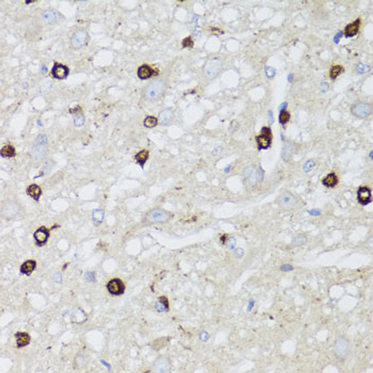
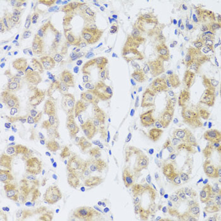
Rabbit polyclonal antibody anti-MIP (50-150) is suitable for use in Western Blot and Immunohistochemistry research applications. |
| Clonality: |
Polyclonal |
| Conjugation: |
Unconjugated |
| Isotype: |
IgG |
| Formulation: |
PBS with 0.02% Sodium Azide, 50% Glycerol, pH7.3. |
| Purification: |
Affinity purification |
| Dilution Range: |
WB 1:500-1:1000IHC-P 1:100-1:200 |
| Storage Instruction: |
Store at-20°C for up to 1 year from the date of receipt, and avoid repeat freeze-thaw cycles. |
| Gene Symbol: |
MIP |
| Gene ID: |
4284 |
| Uniprot ID: |
MIP_HUMAN |
| Immunogen Region: |
50-150 |
| Immunogen: |
A synthetic peptide corresponding to a sequence within amino acids 50-150 of human MIP (NP_036196.1). |
| Immunogen Sequence: |
LALATLVQSVGHISGAHVNP AVTFAFLVGSQMSLLRAFCY MAAQLLGAVAGAAVLYSVTP PAVRGNLALNTLHPAVSVGQ ATTVEIFLTLQFVLCIFATY D |
| Tissue Specificity | Expressed in the cortex and nucleus of the retina lens (at protein level). Major component of lens fiber gap junctions. |
| Post Translational Modifications | Subject to partial proteolytic cleavage in the eye lens core. Partial proteolysis promotes interactions between tetramers from adjoining membranes. Fatty acylated at Met-1 and Lys-238. The acyl modifications, in decreasing order of ion abundance, are: oleoyl (C18:1) > palmitoyl (C16:0) > stearoyl (C18:0) > eicosenoyl (C20:1) > dihomo-gamma-linolenoyl (C20:3) > palmitoleoyl (C16:1) > eicosadienoyl (C20:2). |
| Function | Water channel. Channel activity is down-regulated by CALM when cytoplasmic Ca(2+) levels are increased. May be responsible for regulating the osmolarity of the lens. Interactions between homotetramers from adjoining membranes may stabilize cell junctions in the eye lens core. Plays a role in cell-to-cell adhesion and facilitates gap junction coupling. |
| Protein Name | Lens Fiber Major Intrinsic ProteinAquaporin-0Mip26Mp26 |
| Database Links | Reactome: R-HSA-432047 |
| Cellular Localisation | Cell MembraneMulti-Pass Membrane ProteinCell JunctionGap Junction |
| Alternative Antibody Names | Anti-Lens Fiber Major Intrinsic Protein antibodyAnti-Aquaporin-0 antibodyAnti-Mip26 antibodyAnti-Mp26 antibodyAnti-MIP antibodyAnti-AQP0 antibody |
Information sourced from Uniprot.org
12 months for antibodies. 6 months for ELISA Kits. Please see website T&Cs for further guidance


