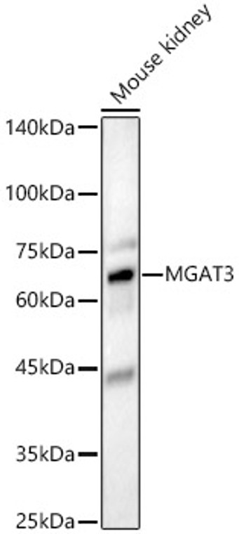
| Host: |
Rabbit |
| Applications: |
WB |
| Reactivity: |
Human/Mouse/Rat |
| Note: |
STRICTLY FOR FURTHER SCIENTIFIC RESEARCH USE ONLY (RUO). MUST NOT TO BE USED IN DIAGNOSTIC OR THERAPEUTIC APPLICATIONS. |
| Short Description: |
Rabbit polyclonal antibody anti-MGAT3 (364-533) is suitable for use in Western Blot research applications. |
| Clonality: |
Polyclonal |
| Conjugation: |
Unconjugated |
| Isotype: |
IgG |
| Formulation: |
PBS with 0.02% Sodium Azide, 50% Glycerol, pH7.3. |
| Purification: |
Affinity purification |
| Dilution Range: |
WB 1:500-1:1000 |
| Storage Instruction: |
Store at-20°C for up to 1 year from the date of receipt, and avoid repeat freeze-thaw cycles. |
| Gene Symbol: |
MGAT3 |
| Gene ID: |
4248 |
| Uniprot ID: |
MGAT3_HUMAN |
| Immunogen Region: |
364-533 |
| Immunogen: |
Recombinant fusion protein containing a sequence corresponding to amino acids 364-533 of human MGAT3 (NP_002400.3). |
| Immunogen Sequence: |
GCTVDMLQAVYGLDGIRLRR RQYYTMPNFRQYENRTGHIL VQWSLGSPLHFAGWHCSWCF TPEGIYFKLVSAQNGDFPRW GDYEDKRDLNYIRGLIRTGG WFDGTQQEYPPADPSEHMYA PKYLLKNYDRFHYLLDNPYQ EPRSTAAGGWRHRGPEGRPP ARGKLDEAEV |
| Function | It is involved in the regulation of the biosynthesis and biological function of glycoprotein oligosaccharides. Catalyzes the addition of N-acetylglucosamine in beta 1-4 linkage to the beta-linked mannose of the trimannosyl core of N-linked sugar chains, called bisecting N-acetylglucosamine (GlcNAc). It is one of the most important enzymes involved in the regulation of the biosynthesis of glycoprotein oligosaccharides. The addition of this bisecting GlcNAc residue alters not only the composition, but also the conformation of the N-glycan. The introduction of the bisecting GlcNAc residue results in the suppression of further processing and elongation of N-glycans, precluding the formation of beta-1,6 GlcNAc branching, catalyzed by MGAT5 since it is unable to use the bisected oligosaccharide as a substrate. Addition of bisecting N-acetylglucosamine to CDH1/E-cadherin modulates CDH1 cell membrane location. Inhibits NeuAc-alpha-2,3-Gal-beta-1,4-GlcNAc- formation which modulates sialylation levels and plays a role in cell migration regulation. In brain, addition of bisecting N-acetylglucosamine to BACE1 blocks its lysosomal targeting in response to oxidative stress and further degradation which increases its location to early endosome and the APP cleavage. |
| Protein Name | Beta-1 -4-Mannosyl-Glycoprotein 4-Beta-N-AcetylglucosaminyltransferaseN-Glycosyl-Oligosaccharide-Glycoprotein N-Acetylglucosaminyltransferase IiiGnt-IiiGlcnac-T IiiN-Acetylglucosaminyltransferase Iii |
| Database Links | Reactome: R-HSA-975574 |
| Cellular Localisation | Golgi Apparatus MembraneSingle-Pass Type Ii Membrane Protein |
| Alternative Antibody Names | Anti-Beta-1 -4-Mannosyl-Glycoprotein 4-Beta-N-Acetylglucosaminyltransferase antibodyAnti-N-Glycosyl-Oligosaccharide-Glycoprotein N-Acetylglucosaminyltransferase Iii antibodyAnti-Gnt-Iii antibodyAnti-Glcnac-T Iii antibodyAnti-N-Acetylglucosaminyltransferase Iii antibodyAnti-MGAT3 antibodyAnti-GGNT3 antibody |
Information sourced from Uniprot.org
12 months for antibodies. 6 months for ELISA Kits. Please see website T&Cs for further guidance


