| Host: |
Rabbit |
| Applications: |
IF |
| Reactivity: |
Human |
| Note: |
STRICTLY FOR FURTHER SCIENTIFIC RESEARCH USE ONLY (RUO). MUST NOT TO BE USED IN DIAGNOSTIC OR THERAPEUTIC APPLICATIONS. |
| Short Description: |
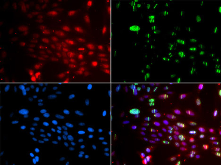
Rabbit polyclonal antibody anti-L3MBTL1 (533-772) is suitable for use in Immunofluorescence research applications. |
| Clonality: |
Polyclonal |
| Conjugation: |
Unconjugated |
| Isotype: |
IgG |
| Formulation: |
PBS with 0.02% Sodium Azide, 50% Glycerol, pH7.3. |
| Purification: |
Affinity purification |
| Dilution Range: |
IF/ICC 1:50-1:100 |
| Storage Instruction: |
Store at-20°C for up to 1 year from the date of receipt, and avoid repeat freeze-thaw cycles. |
| Gene Symbol: |
L3MBTL1 |
| Gene ID: |
26013 |
| Uniprot ID: |
LMBL1_HUMAN |
| Immunogen Region: |
533-772 |
| Immunogen: |
Recombinant fusion protein containing a sequence corresponding to amino acids 533-772 of human L3MBTL1 (NP_056293.4). |
| Immunogen Sequence: |
PLSYRSLPHTRTSKYSFHHR KCPTPGCDGSGHVTGKFTAH HCLSGCPLAERNQSRLKAEL SDSEASARKKNLSGFSPRKK PRHHGRIGRPPKYRKIPQED FQTLTPDVVHQSLFMSALSA HPDRSLSVCWEQHCKLLPGV AGISASTVAKWTIDEVFGFV QTLTGCEDQARLFKDEARIV RVTHVSGKTLVWTVAQLGDL VCSDHLQEGKGILETGVHSL LCSLPTHLLAKLSFASDSQ |
| Tissue Specificity | Widely expressed. Expression is reduced in colorectal cancer cell line SW480 and promyelocytic leukemia cell line HL-60. |
| Post Translational Modifications | Ubiquitinated in a VCP/p97-dependent way following DNA damage, leading to its removal from DNA damage sites, promoting accessibility of H4K20me2 mark for DNA repair protein TP53BP1, which is then recruited to DNA damage sites. |
| Function | Polycomb group (PcG) protein that specifically recognizes and binds mono- and dimethyllysine residues on target proteins, therey acting as a 'reader' of a network of post-translational modifications. PcG proteins maintain the transcriptionally repressive state of genes: acts as a chromatin compaction factor by recognizing and binding mono- and dimethylated histone H1b/H1-4 at 'Lys-26' (H1bK26me1 and H1bK26me2) and histone H4 at 'Lys-20' (H4K20me1 and H4K20me2), leading to condense chromatin and repress transcription. Recognizes and binds p53/TP53 monomethylated at 'Lys-382', leading to repress p53/TP53-target genes. Also recognizes and binds RB1/RB monomethylated at 'Lys-860'. Participates in the ETV6-mediated repression. Probably plays a role in cell proliferation. Overexpression induces multinucleated cells, suggesting that it is required to accomplish normal mitosis. |
| Protein Name | Lethal(3malignant Brain Tumor-Like Protein 1H-L(3mbtH-L(3mbt ProteinL(3mbt-LikeL(3mbt Protein HomologL3mbtl1 |
| Database Links | Reactome: R-HSA-6804760 |
| Cellular Localisation | NucleusExcluded From The NucleolusDoes Not Colocalize With The Pcg Protein Bmi1Suggesting That These Two Proteins Do Not Belong To The Same Complex |
| Alternative Antibody Names | Anti-Lethal(3malignant Brain Tumor-Like Protein 1 antibodyAnti-H-L(3mbt antibodyAnti-H-L(3mbt Protein antibodyAnti-L(3mbt-Like antibodyAnti-L(3mbt Protein Homolog antibodyAnti-L3mbtl1 antibodyAnti-L3MBTL1 antibodyAnti-KIAA0681 antibodyAnti-L3MBT antibodyAnti-L3MBTL antibody |
Information sourced from Uniprot.org
12 months for antibodies. 6 months for ELISA Kits. Please see website T&Cs for further guidance


