| Host: |
Rabbit |
| Applications: |
WB/IHC/IF |
| Reactivity: |
Human/Mouse/Rat |
| Note: |
STRICTLY FOR FURTHER SCIENTIFIC RESEARCH USE ONLY (RUO). MUST NOT TO BE USED IN DIAGNOSTIC OR THERAPEUTIC APPLICATIONS. |
| Short Description: |
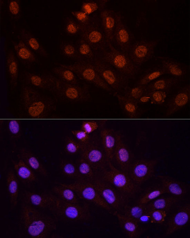
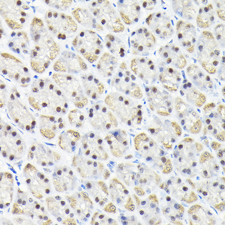
Rabbit polyclonal antibody anti-KPNA3 (1-210) is suitable for use in Western Blot, Immunohistochemistry and Immunofluorescence research applications. |
| Clonality: |
Polyclonal |
| Conjugation: |
Unconjugated |
| Isotype: |
IgG |
| Formulation: |
PBS with 0.02% Sodium Azide, 50% Glycerol, pH7.3. |
| Purification: |
Affinity purification |
| Dilution Range: |
WB 1:500-1:2000IHC-P 1:50-1:200IF/ICC 1:50-1:200 |
| Storage Instruction: |
Store at-20°C for up to 1 year from the date of receipt, and avoid repeat freeze-thaw cycles. |
| Gene Symbol: |
KPNA3 |
| Gene ID: |
3839 |
| Uniprot ID: |
IMA4_HUMAN |
| Immunogen Region: |
1-210 |
| Immunogen: |
Recombinant fusion protein containing a sequence corresponding to amino acids 1-210 of human KPNA3 (NP_002258.2). |
| Immunogen Sequence: |
MAENPSLENHRIKSFKNKGR DVETMRRHRNEVTVELRKNK RDEHLLKKRNVPQEESLEDS DVDADFKAQNVTLEAILQNA TSDNPVVQLSAVQAARKLLS SDRNPPIDDLIKSGILPILV KCLERDDNPSLQFEAAWALT NIASGTSAQTQAVVQSNAVP LFLRLLRSPHQNVCEQAVWA LGNIIGDGPQCRDYVISLGV VKPLLSFISP |
| Tissue Specificity | Ubiquitous. Highest levels in heart and skeletal muscle. |
| Function | Functions in nuclear protein import as an adapter protein for nuclear receptor KPNB1. Binds specifically and directly to substrates containing either a simple or bipartite NLS motif. Docking of the importin/substrate complex to the nuclear pore complex (NPC) is mediated by KPNB1 through binding to nucleoporin FxFG repeats and the complex is subsequently translocated through the pore by an energy requiring, Ran-dependent mechanism. At the nucleoplasmic side of the NPC, Ran binds to importin-beta and the three components separate and importin-alpha and -beta are re-exported from the nucleus to the cytoplasm where GTP hydrolysis releases Ran from importin. The directionality of nuclear import is thought to be conferred by an asymmetric distribution of the GTP- and GDP-bound forms of Ran between the cytoplasm and nucleus. In vitro, mediates the nuclear import of human cytomegalovirus UL84 by recognizing a non-classical NLS. Recognizes NLSs of influenza A virus nucleoprotein probably through ARM repeats 7-9. |
| Protein Name | Importin Subunit Alpha-4Importin Alpha Q2Qip2Karyopherin Subunit Alpha-3Srp1-Gamma |
| Database Links | Reactome: R-HSA-1169408Reactome: R-HSA-168276 |
| Cellular Localisation | CytoplasmNucleus |
| Alternative Antibody Names | Anti-Importin Subunit Alpha-4 antibodyAnti-Importin Alpha Q2 antibodyAnti-Qip2 antibodyAnti-Karyopherin Subunit Alpha-3 antibodyAnti-Srp1-Gamma antibodyAnti-KPNA3 antibodyAnti-QIP2 antibody |
Information sourced from Uniprot.org
12 months for antibodies. 6 months for ELISA Kits. Please see website T&Cs for further guidance


