| Host: |
Rabbit |
| Applications: |
WB |
| Reactivity: |
Mouse/Rat |
| Note: |
STRICTLY FOR FURTHER SCIENTIFIC RESEARCH USE ONLY (RUO). MUST NOT TO BE USED IN DIAGNOSTIC OR THERAPEUTIC APPLICATIONS. |
| Short Description: |
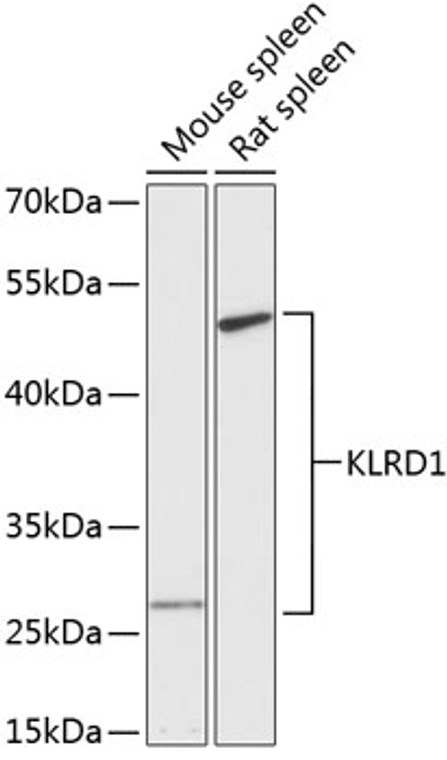
Rabbit polyclonal antibody anti-KLRD1 (32-179) is suitable for use in Western Blot research applications. |
| Clonality: |
Polyclonal |
| Conjugation: |
Unconjugated |
| Isotype: |
IgG |
| Formulation: |
PBS with 0.01% Thimerosal, 50% Glycerol, pH7.3. |
| Purification: |
Affinity purification |
| Dilution Range: |
WB 1:500-1:2000 |
| Storage Instruction: |
Store at-20°C for up to 1 year from the date of receipt, and avoid repeat freeze-thaw cycles. |
| Gene Symbol: |
KLRD1 |
| Gene ID: |
3824 |
| Uniprot ID: |
KLRD1_HUMAN |
| Immunogen Region: |
32-179 |
| Immunogen: |
Recombinant fusion protein containing a sequence corresponding to amino acids 32-179 of human KLRD1 (NP_002253.2). |
| Immunogen Sequence: |
KNSFTKLSIEPAFTPGPNIE LQKDSDCCSCQEKWVGYRCN CYFISSEQKTWNESRHLCAS QKSSLLQLQNTDELDFMSSS QQFYWIGLSYSEEHTAWLWE NGSALSQYLFPSFETFNTKN CIAYNPNGNALDESCEDKNR YICKQQLI |
| Tissue Specificity | Expressed in NK cell subsets (at protein level). Expressed in memory/effector CD8-positive alpha-beta T cell subsets (at protein level). Expressed in melanoma-specific cytotoxic T cell clones (at protein level). Expressed in terminally differentiated cytotoxic gamma-delta T cells (at protein level). KLRD1-KLRC1 and KLRD1-KLRC2 are differentially expressed in NK and T cell populations, with only minor subsets expressing both receptor complexes (at protein level). |
| Function | Immune receptor involved in self-nonself discrimination. In complex with KLRC1 or KLRC2 on cytotoxic and regulatory lymphocyte subsets, recognizes non-classical major histocompatibility (MHC) class Ib molecule HLA-E loaded with self-peptides derived from the signal sequence of classical MHC class Ia and non-classical MHC class Ib molecules. Enables cytotoxic cells to monitor the expression of MHC class I molecules in healthy cells and to tolerate self. Primarily functions as a ligand binding subunit as it lacks the capacity to signal. KLRD1-KLRC1 acts as an immune inhibitory receptor. Key inhibitory receptor on natural killer (NK) cells that regulates their activation and effector functions. Dominantly counteracts T cell receptor signaling on a subset of memory/effector CD8-positive T cells as part of an antigen-driven response to avoid autoimmunity. On intraepithelial CD8-positive gamma-delta regulatory T cells triggers TGFB1 secretion, which in turn limits the cytotoxic programming of intraepithelial CD8-positive alpha-beta T cells, distinguishing harmless from pathogenic antigens. In HLA-E-rich tumor microenvironment, acts as an immune inhibitory checkpoint and may contribute to progressive loss of effector functions of NK cells and tumor-specific T cells, a state known as cell exhaustion. Upon HLA-E-peptide binding, transmits intracellular signals through KLRC1 immunoreceptor tyrosine-based inhibition motifs (ITIMs) by recruiting INPP5D/SHIP-1 and INPPL1/SHIP-2 tyrosine phosphatases to ITIMs, and ultimately opposing signals transmitted by activating receptors through dephosphorylation of proximal signaling molecules. KLRD1-KLRC2 acts as an immune activating receptor. On cytotoxic lymphocyte subsets recognizes HLA-E loaded with signal sequence-derived peptides from non-classical MHC class Ib HLA-G molecules, likely playing a role in the generation and effector functions of adaptive NK cells and in maternal-fetal tolerance during pregnancy. Regulates the effector functions of terminally differentiated cytotoxic lymphocyte subsets, and in particular may play a role in adaptive NK cell response to viral infection. Upon HLA-E-peptide binding, transmits intracellular signals via the adapter protein TYROBP/DAP12, triggering the phosphorylation of proximal signaling molecules and cell activation. (Microbial infection) Viruses like human cytomegalovirus have evolved an escape mechanism whereby virus-induced down-regulation of host MHC class I molecules is coupled to the binding of viral peptides to HLA-E, restoring HLA-E expression and inducing HLA-E-dependent NK cell immune tolerance to infected cells. Recognizes HLA-E in complex with human cytomegalovirus UL40-derived peptide (VMAPRTLIL) and inhibits NK cell cytotoxicity. (Microbial infection) May recognize HLA-E in complex with HIV-1 gag/Capsid protein p24-derived peptide (AISPRTLNA) on infected cells and may inhibit NK cell cytotoxicity, a mechanism that allows HIV-1 to escape immune recognition. (Microbial infection) Upon SARS-CoV-2 infection, may contribute to functional exhaustion of cytotoxic NK cells and CD8-positive T cells. On NK cells, may recognize HLA-E in complex with SARS-CoV-2 S/Spike protein S1-derived peptide (LQPRTFLL) expressed on the surface of lung epithelial cells, inducing NK cell exhaustion and dampening antiviral immune surveillance. |
| Protein Name | Natural Killer Cells Antigen Cd94Kp43Killer Cell Lectin-Like Receptor Subfamily D Member 1Nk Cell ReceptorCd Antigen Cd94 |
| Database Links | Reactome: R-HSA-198933Reactome: R-HSA-2172127Reactome: R-HSA-2424491 |
| Cellular Localisation | Cell MembraneSingle-Pass Type Ii Membrane Protein |
| Alternative Antibody Names | Anti-Natural Killer Cells Antigen Cd94 antibodyAnti-Kp43 antibodyAnti-Killer Cell Lectin-Like Receptor Subfamily D Member 1 antibodyAnti-Nk Cell Receptor antibodyAnti-Cd Antigen Cd94 antibodyAnti-KLRD1 antibodyAnti-CD94 antibody |
Information sourced from Uniprot.org
12 months for antibodies. 6 months for ELISA Kits. Please see website T&Cs for further guidance


