| Host: |
Rabbit |
| Applications: |
WB |
| Reactivity: |
Human/Mouse |
| Note: |
STRICTLY FOR FURTHER SCIENTIFIC RESEARCH USE ONLY (RUO). MUST NOT TO BE USED IN DIAGNOSTIC OR THERAPEUTIC APPLICATIONS. |
| Short Description: |
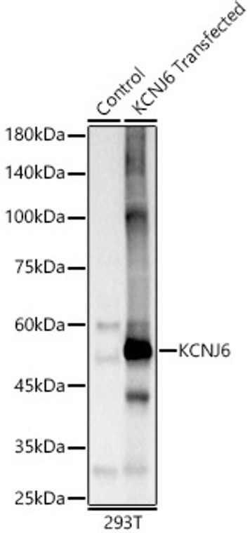
Rabbit polyclonal antibody anti-KCNJ6 (1-80) is suitable for use in Western Blot research applications. |
| Clonality: |
Polyclonal |
| Conjugation: |
Unconjugated |
| Isotype: |
IgG |
| Formulation: |
PBS with 0.02% Sodium Azide, 50% Glycerol, pH7.3. |
| Purification: |
Affinity purification |
| Dilution Range: |
WB 1:500-1:2000 |
| Storage Instruction: |
Store at-20°C for up to 1 year from the date of receipt, and avoid repeat freeze-thaw cycles. |
| Gene Symbol: |
KCNJ6 |
| Gene ID: |
3763 |
| Uniprot ID: |
KCNJ6_HUMAN |
| Immunogen Region: |
1-80 |
| Immunogen: |
Recombinant fusion protein containing a sequence corresponding to amino acids 1-80 of human KCNJ6 (NP_002231.1). |
| Immunogen Sequence: |
MAKLTESMTNVLEGDSMDQD VESPVAIHQPKLPKQARDDL PRHISRDRTKRKIQRYVRKD GKCNVHHGNVRETYRYLTDI |
| Tissue Specificity | Most abundant in cerebellum, and to a lesser degree in islets and exocrine pancreas. |
| Function | This potassium channel may be involved in the regulation of insulin secretion by glucose and/or neurotransmitters acting through G-protein-coupled receptors. Inward rectifier potassium channels are characterized by a greater tendency to allow potassium to flow into the cell rather than out of it. Their voltage dependence is regulated by the concentration of extracellular potassium.as external potassium is raised, the voltage range of the channel opening shifts to more positive voltages. The inward rectification is mainly due to the blockage of outward current by internal magnesium. |
| Protein Name | G Protein-Activated Inward Rectifier Potassium Channel 2Girk-2Bir1Inward Rectifier K(+ Channel Kir3.2Katp-2Potassium Channel - Inwardly Rectifying Subfamily J Member 6 |
| Database Links | Reactome: R-HSA-1296041Reactome: R-HSA-997272 |
| Cellular Localisation | MembraneMulti-Pass Membrane Protein |
| Alternative Antibody Names | Anti-G Protein-Activated Inward Rectifier Potassium Channel 2 antibodyAnti-Girk-2 antibodyAnti-Bir1 antibodyAnti-Inward Rectifier K(+ Channel Kir3.2 antibodyAnti-Katp-2 antibodyAnti-Potassium Channel - Inwardly Rectifying Subfamily J Member 6 antibodyAnti-KCNJ6 antibodyAnti-GIRK2 antibodyAnti-KATP2 antibodyAnti-KCNJ7 antibody |
Information sourced from Uniprot.org
12 months for antibodies. 6 months for ELISA Kits. Please see website T&Cs for further guidance


