| Host: |
Rabbit |
| Applications: |
WB |
| Reactivity: |
Human/Mouse |
| Note: |
STRICTLY FOR FURTHER SCIENTIFIC RESEARCH USE ONLY (RUO). MUST NOT TO BE USED IN DIAGNOSTIC OR THERAPEUTIC APPLICATIONS. |
| Short Description: |
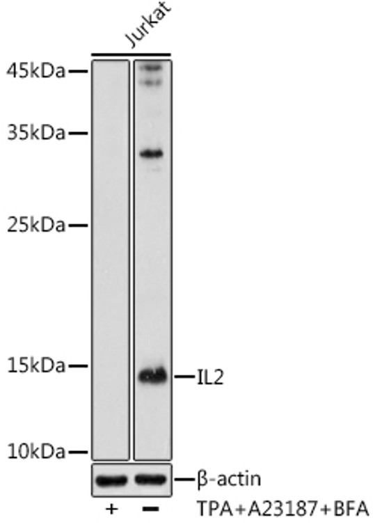
Rabbit polyclonal antibody anti-IL2 (21-153) is suitable for use in Western Blot research applications. |
| Clonality: |
Polyclonal |
| Conjugation: |
Unconjugated |
| Isotype: |
IgG |
| Formulation: |
PBS with 0.01% Thimerosal, 50% Glycerol, pH7.3. |
| Purification: |
Affinity purification |
| Dilution Range: |
WB 1:500-1:1000 |
| Storage Instruction: |
Store at-20°C for up to 1 year from the date of receipt, and avoid repeat freeze-thaw cycles. |
| Gene Symbol: |
IL2 |
| Gene ID: |
3558 |
| Uniprot ID: |
IL2_HUMAN |
| Immunogen Region: |
21-153 |
| Immunogen: |
Recombinant fusion protein containing a sequence corresponding to amino acids 21-153 of IL2 (NP_000577.2). |
| Immunogen Sequence: |
APTSSSTKKTQLQLEHLLLD LQMILNGINNYKNPKLTRML TFKFYMPKKATELKHLQCLE EELKPLEEVLNLAQSKNFHL RPRDLISNINVIVLELKGSE TTFMCEYADETATIVEFLNR WITFCQSIISTLT |
| Function | Cytokine produced by activated CD4-positive helper T-cells and to a lesser extend activated CD8-positive T-cells and natural killer (NK) cells that plays pivotal roles in the immune response and tolerance. Binds to a receptor complex composed of either the high-affinity trimeric IL-2R (IL2RA/CD25, IL2RB/CD122 and IL2RG/CD132) or the low-affinity dimeric IL-2R (IL2RB and IL2RG). Interaction with the receptor leads to oligomerization and conformation changes in the IL-2R subunits resulting in downstream signaling starting with phosphorylation of JAK1 and JAK3. In turn, JAK1 and JAK3 phosphorylate the receptor to form a docking site leading to the phosphorylation of several substrates including STAT5. This process leads to activation of several pathways including STAT, phosphoinositide-3-kinase/PI3K and mitogen-activated protein kinase/MAPK pathways. Functions as a T-cell growth factor and can increase NK-cell cytolytic activity as well. Promotes strong proliferation of activated B-cells and subsequently immunoglobulin production. Plays a pivotal role in regulating the adaptive immune system by controlling the survival and proliferation of regulatory T-cells, which are required for the maintenance of immune tolerance. Moreover, participates in the differentiation and homeostasis of effector T-cell subsets, including Th1, Th2, Th17 as well as memory CD8-positive T-cells. |
| Protein Name | Interleukin-2Il-2T-Cell Growth FactorTcgfAldesleukin |
| Database Links | Reactome: R-HSA-5673001Reactome: R-HSA-8877330Reactome: R-HSA-9020558Reactome: R-HSA-912526 |
| Cellular Localisation | Secreted |
| Alternative Antibody Names | Anti-Interleukin-2 antibodyAnti-Il-2 antibodyAnti-T-Cell Growth Factor antibodyAnti-Tcgf antibodyAnti-Aldesleukin antibodyAnti-IL2 antibody |
Information sourced from Uniprot.org
12 months for antibodies. 6 months for ELISA Kits. Please see website T&Cs for further guidance


