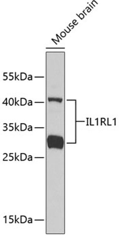
| Host: |
Rabbit |
| Applications: |
WB |
| Reactivity: |
Human/Mouse/Rat |
| Note: |
STRICTLY FOR FURTHER SCIENTIFIC RESEARCH USE ONLY (RUO). MUST NOT TO BE USED IN DIAGNOSTIC OR THERAPEUTIC APPLICATIONS. |
| Short Description: |
Rabbit polyclonal antibody anti-IL1RL1 (19-328) is suitable for use in Western Blot research applications. |
| Clonality: |
Polyclonal |
| Conjugation: |
Unconjugated |
| Isotype: |
IgG |
| Formulation: |
PBS with 0.05% Proclin300, 50% Glycerol, pH7.3. |
| Purification: |
Affinity purification |
| Dilution Range: |
WB 1:500-1:2000 |
| Storage Instruction: |
Store at-20°C for up to 1 year from the date of receipt, and avoid repeat freeze-thaw cycles. |
| Gene Symbol: |
IL1RL1 |
| Gene ID: |
9173 |
| Uniprot ID: |
ILRL1_HUMAN |
| Immunogen Region: |
19-328 |
| Immunogen: |
Recombinant fusion protein containing a sequence corresponding to amino acids 19-328 of human IL1RL1 (NP_057316.3). |
| Immunogen Sequence: |
KFSKQSWGLENEALIVRCPR QGKPSYTVDWYYSQTNKSIP TQERNRVFASGQLLKFLPAA VADSGIYTCIVRSPTFNRTG YANVTIYKKQSDCNVPDYLM YSTVSGSEKNSKIYCPTIDL YNWTAPLEWFKNCQALQGSR YRAHKSFLVIDNVMTEDAGD YTCKFIHNENGANYSVTATR SFTVKDEQGFSLFPVIGAPA QNEIKEVEIGKNANLTCSAC FGKGTQFLAAVLWQLNGTK |
| Tissue Specificity | Highly expressed in kidney, lung, placenta, stomach, skeletal muscle, colon and small intestine. Isoform A is prevalently expressed in the lung, testis, placenta, stomach and colon. Isoform B is more abundant in the brain, kidney and the liver. Isoform C is not detected in brain, heart, liver, kidney and skeletal muscle. Expressed on T-cells in fibrotic liver.at protein level. Overexpressed in fibrotic and cirrhotic liver. |
| Post Translational Modifications | Ubiquitinated at Lys-321 in a FBXL19-mediated manner.leading to proteasomal degradation. Ubiquitination by TRAF6 via 'Lys-27'-linked polyubiquitination and deubiquitination by USP38 serves as a critical regulatory mechanism for fine-tuning IL1RL1-mediated inflammatory response. |
| Function | Receptor for interleukin-33 (IL-33) which plays crucial roles in innate and adaptive immunity, contributing to tissue homeostasis and responses to environmental stresses together with coreceptor IL1RAP. Its stimulation recruits MYD88, IRAK1, IRAK4, and TRAF6, followed by phosphorylation of MAPK3/ERK1 and/or MAPK1/ERK2, MAPK14, and MAPK8. Possibly involved in helper T-cell function (Probable). Upon tissue injury, induces UCP2-dependent mitochondrial rewiring that attenuates the generation of reactive oxygen species and preserves the integrity of Krebs cycle required for persistent production of itaconate and subsequent GATA3-dependent differentiation of inflammation-resolving alternatively activated macrophages. Isoform B: Inhibits IL-33 signaling. |
| Protein Name | Interleukin-1 Receptor-Like 1Protein St2 |
| Database Links | Reactome: R-HSA-1257604Reactome: R-HSA-6811558Reactome: R-HSA-9014843 Q01638-2 |
| Cellular Localisation | Isoform C: Cell MembraneIsoform B: SecretedCell MembraneSingle-Pass Type I Membrane Protein |
| Alternative Antibody Names | Anti-Interleukin-1 Receptor-Like 1 antibodyAnti-Protein St2 antibodyAnti-IL1RL1 antibodyAnti-DER4 antibodyAnti-ST2 antibodyAnti-T1 antibody |
Information sourced from Uniprot.org
12 months for antibodies. 6 months for ELISA Kits. Please see website T&Cs for further guidance


