| Host: |
Rabbit |
| Applications: |
WB/IHC/IF/IP |
| Reactivity: |
Human/Mouse |
| Note: |
STRICTLY FOR FURTHER SCIENTIFIC RESEARCH USE ONLY (RUO). MUST NOT TO BE USED IN DIAGNOSTIC OR THERAPEUTIC APPLICATIONS. |
| Short Description: |
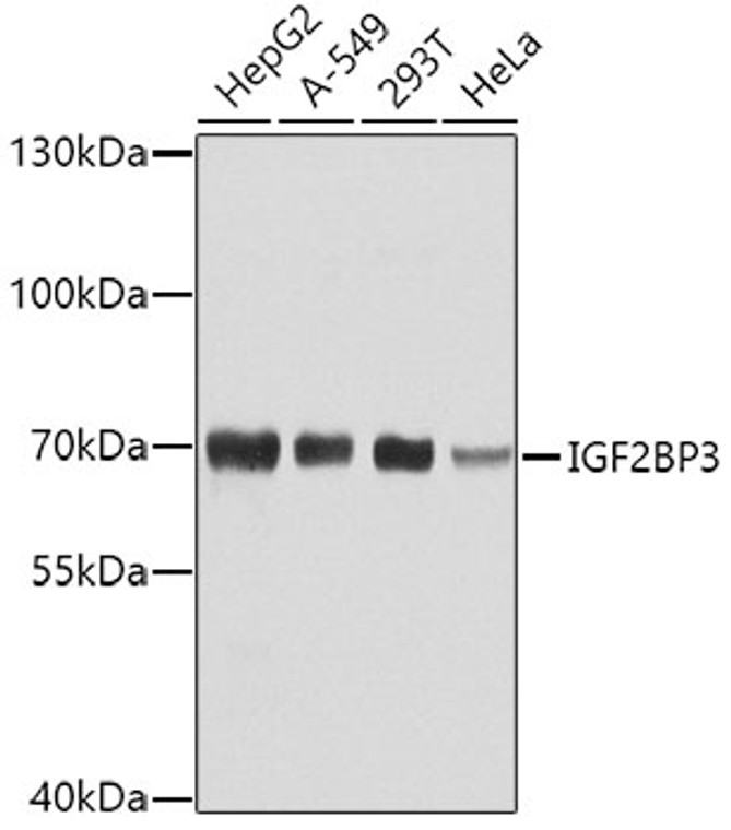
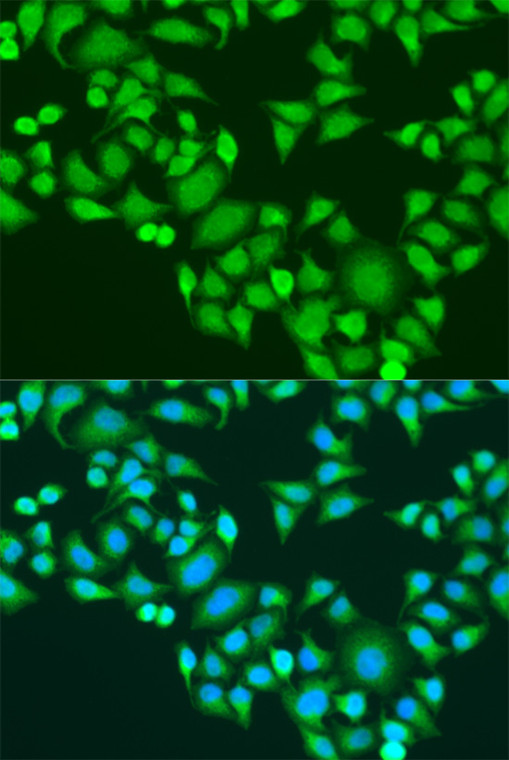
Rabbit polyclonal antibody anti-IGF2BP3 (300-579) is suitable for use in Western Blot, Immunohistochemistry, Immunofluorescence and Immunoprecipitation research applications. |
| Clonality: |
Polyclonal |
| Conjugation: |
Unconjugated |
| Isotype: |
IgG |
| Formulation: |
PBS with 0.02% Sodium Azide, 50% Glycerol, pH7.3. |
| Purification: |
Affinity purification |
| Dilution Range: |
WB 1:500-1:2000IHC-P 1:50-1:200IF/ICC 1:50-1:200IP 1:50-1:200 |
| Storage Instruction: |
Store at-20°C for up to 1 year from the date of receipt, and avoid repeat freeze-thaw cycles. |
| Gene Symbol: |
IGF2BP3 |
| Gene ID: |
10643 |
| Uniprot ID: |
IF2B3_HUMAN |
| Immunogen Region: |
300-579 |
| Immunogen: |
A synthetic peptide corresponding to a sequence within amino acids 300-579 of human IGF2BP3 (NP_006538.2). |
| Immunogen Sequence: |
KKIEQDTDTKITISPLQELT LYNPERTITVKGNVETCAKA EEEIMKKIRESYENDIASMN LQAHLIPGLNLNALGLFPPT SGMPPPTSGPPSAMTPPYPQ FEQSETETVHLFIPALSVGA IIGKQGQHIKQLSRFAGASI KIAPAEAPDAKVRMVIITGP PEAQFKAQGRIYGKIKEENF VSPKEEVKLEAHIRVPSFAA GRVIGKGGKTVNELQNLSSA EVVVPRDQTPDENDQVVVK |
| Tissue Specificity | Expressed in fetal liver, fetal lung, fetal kidney, fetal thymus, fetal placenta, fetal follicles of ovary and gonocytes of testis, growing oocytes, spermatogonia and semen (at protein level). Expressed in cervix adenocarcinoma, in testicular, pancreatic and renal-cell carcinomas (at protein level). Expressed ubiquitously during fetal development at 8 and 14 weeks of gestation. Expressed in ovary, testis, brain, placenta, pancreatic cancer tissues and pancreatic cancer cell lines. |
| Function | RNA-binding factor that may recruit target transcripts to cytoplasmic protein-RNA complexes (mRNPs). This transcript 'caging' into mRNPs allows mRNA transport and transient storage. It also modulates the rate and location at which target transcripts encounter the translational apparatus and shields them from endonuclease attacks or microRNA-mediated degradation. Preferentially binds to N6-methyladenosine (m6A)-containing mRNAs and increases their stability. Binds to the 3'-UTR of CD44 mRNA and stabilizes it, hence promotes cell adhesion and invadopodia formation in cancer cells. Binds to beta-actin/ACTB and MYC transcripts. Increases MYC mRNA stability by binding to the coding region instability determinant (CRD) and binding is enhanced by m6A-modification of the CRD. Binds to the 5'-UTR of the insulin-like growth factor 2 (IGF2) mRNAs. |
| Protein Name | Insulin-Like Growth Factor 2 Mrna-Binding Protein 3Igf2 Mrna-Binding Protein 3Imp-3Igf-Ii Mrna-Binding Protein 3Kh Domain-Containing Protein Overexpressed In CancerHkocVickz Family Member 3 |
| Database Links | Reactome: R-HSA-428359 |
| Cellular Localisation | NucleusCytoplasmP-BodyStress GranuleFound In Lamellipodia Of The Leading EdgeIn The Perinuclear RegionAnd Beneath The Plasma MembraneThe Subcytoplasmic Localization Is Cell Specific And Regulated By Cell Contact And GrowthLocalized At The Connecting Piece And The Tail Of The SpermatozoaColocalized With Cd44 Mrna In Rnp GranulesIn Response To Cellular StressSuch As Oxidative StressRecruited To Stress Granules |
| Alternative Antibody Names | Anti-Insulin-Like Growth Factor 2 Mrna-Binding Protein 3 antibodyAnti-Igf2 Mrna-Binding Protein 3 antibodyAnti-Imp-3 antibodyAnti-Igf-Ii Mrna-Binding Protein 3 antibodyAnti-Kh Domain-Containing Protein Overexpressed In Cancer antibodyAnti-Hkoc antibodyAnti-Vickz Family Member 3 antibodyAnti-IGF2BP3 antibodyAnti-IMP3 antibodyAnti-KOC1 antibodyAnti-VICKZ3 antibody |
Information sourced from Uniprot.org
12 months for antibodies. 6 months for ELISA Kits. Please see website T&Cs for further guidance


