• Western blot analysis of extracts of various cell lines, using HSP20/HSPB6 rabbit monoclonal antibody (STJ11102422) at 1:1000 dilution. Secondary antibody: HRP Goat Anti-rabbit IgG (H+L) at 1:10000 dilution. Lysates/proteins: 25ug per lane. Blocking buffer: 3% non-fat dry milk in TBST. Detection: ECL Basic Kit. Exposure time: 1s.
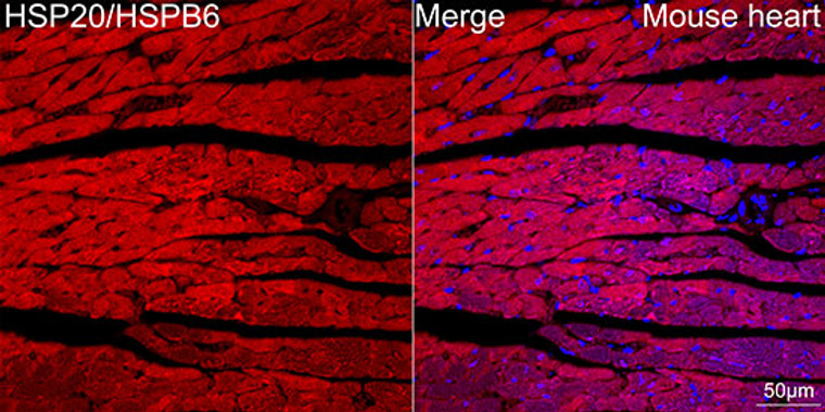
  • Confocal imaging of paraffin-embedded Mouse heart tissue using HSP20/HSPB6 Rabbit monoclonal antibody (STJ11102422, dilution 1:200) followed by a further incubation with Cy3 Goat Anti-Rabbit IgG (H+L) ( dilution 1:500) (Red). DAPI was used for nuclear staining (Blue). Objective: 40x. Perform high pressure antigen retrieval with 0. 01 M citrate buffer (pH 6. 0) prior to IF staining.
  • Immunohistochemistry of paraffin-embedded rat heart using HSP20/HSPB6 rabbit monoclonal antibody (STJ11102422) at dilution of 1:100 (40x lens).
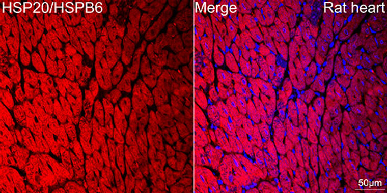
  • Confocal imaging of paraffin-embedded Rat heart tissue using HSP20/HSPB6 Rabbit monoclonal antibody (STJ11102422, dilution 1:200) followed by a further incubation with Cy3 Goat Anti-Rabbit IgG (H+L) ( dilution 1:500) (Red). DAPI was used for nuclear staining (Blue). Objective: 40x. Perform high pressure antigen retrieval with 0. 01 M citrate buffer (pH 6. 0) prior to IF staining.
  • Immunohistochemistry of paraffin-embedded human liver using HSP20/HSPB6 rabbit monoclonal antibody (STJ11102422) at dilution of 1:100 (40x lens).
  • Immunohistochemistry analysis of HSP20/HSPB6 in paraffin-embedded rat heart tissue using HSP20/HSPB6 Rabbit monoclonal antibody (STJ11102422) at a dilution of 1:200 (40x lens). High pressure antigen retrieval was performed with 0. 01 M Tris-EDTA buffer (pH 9. 0) prior to immunohistochemistry staining.
  • Immunohistochemistry of paraffin-embedded mouse heart using HSP20/HSPB6 rabbit monoclonal antibody (STJ11102422) at dilution of 1:100 (40x lens).
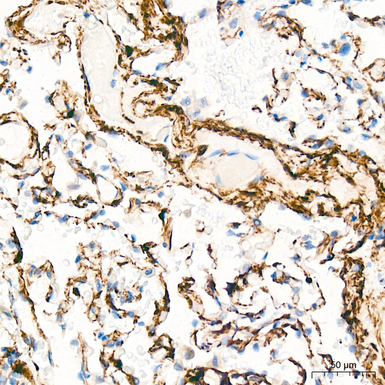
  • Immunohistochemistry analysis of HSP20/HSPB6 in paraffin-embedded human lung tissue using HSP20/HSPB6 Rabbit monoclonal antibody (STJ11102422) at a dilution of 1:200 (40x lens). High pressure antigen retrieval was performed with 0. 01 M Tris-EDTA buffer (pH 9. 0) prior to immunohistochemistry staining.
  • Immunofluorescence analysis of rat heart using HSP20/HSPB6 rabbit monoclonal antibody (STJ11102422) at dilution of 1:100 (40x lens). Blue: DAPI for nuclear staining.
  • Immunohistochemistry analysis of HSP20/HSPB6 in paraffin-embedded rat colon tissue using HSP20/HSPB6 Rabbit monoclonal antibody (STJ11102422) at a dilution of 1:200 (40x lens). High pressure antigen retrieval was performed with 0. 01 M Tris-EDTA buffer (pH 9. 0) prior to immunohistochemistry staining.
  • Immunohistochemistry analysis of HSP20/HSPB6 in paraffin-embedded human liver cancer tissue using HSP20/HSPB6 Rabbit monoclonal antibody (STJ11102422) at a dilution of 1:200 (40x lens). High pressure antigen retrieval was performed with 0. 01 M Tris-EDTA buffer (pH 9. 0) prior to immunohistochemistry staining.
  • Immunohistochemistry analysis of HSP20/HSPB6 in paraffin-embedded human colon carcinoma tissue using HSP20/HSPB6 Rabbit monoclonal antibody (STJ11102422) at a dilution of 1:200 (40x lens). High pressure antigen retrieval was performed with 0. 01 M Tris-EDTA buffer (pH 9. 0) prior to immunohistochemistry staining.
  • Immunohistochemistry analysis of HSP20/HSPB6 in paraffin-embedded mouse heart tissue using HSP20/HSPB6 Rabbit monoclonal antibody (STJ11102422) at a dilution of 1:200 (40x lens). High pressure antigen retrieval was performed with 0. 01 M Tris-EDTA buffer (pH 9. 0) prior to immunohistochemistry staining.
  • Western blot analysis of various lysates using HSP20/HSPB6 Rabbit monoclonal antibody (STJ11102422) at 1:1000 dilution. Secondary antibody: HRP Goat Anti-Rabbit IgG (H+L) (STJS000856) at 1:10000 dilution. Lysates/proteins: 25 Mu g per lane. Blocking buffer: 3% nonfat dry milk in TBST. Detection: ECL Basic Kit. Exposure time: 1s.

Anti-HSPB6 antibody (70-150) [S2MR] (STJ11102422)

SKU:
STJ11102422

Shipping:
Free Shipping
Current Stock:
Host: Rabbit
Applications: WB/IHC/IF
Reactivity: Human/Mouse/Rat
Note: STRICTLY FOR FURTHER SCIENTIFIC RESEARCH USE ONLY (RUO). MUST NOT TO BE USED IN DIAGNOSTIC OR THERAPEUTIC APPLICATIONS.
Short Description: Rabbit monoclonal antibody anti-Hsp20 (70-150) is suitable for use in Western Blot, Immunohistochemistry and Immunofluorescence research applications.
Clonality: Monoclonal
Clone ID: S2MR
Conjugation: Unconjugated
Isotype: IgG
Formulation: PBS with 0.02% Sodium Azide, 0.05% BSA, 50% Glycerol, pH7.3.
Purification: Affinity purification
Dilution Range: WB 1:500-1:1000
IHC-P 1:50-1:200
IF/ICC 1:50-1:200
Storage Instruction: Store at-20°C for up to 1 year from the date of receipt, and avoid repeat freeze-thaw cycles.
Gene Symbol: HSPB6
Gene ID: 126393
Uniprot ID: HSPB6_HUMAN
Immunogen Region: 70-150
Immunogen: A synthetic peptide corresponding to a sequence within amino acids 70-150 of human HSP20/HSPB6 (O14558).
Immunogen Sequence: DPGHFSVLLDVKHFSPEEIA VKVVGEHVEVHARHEERPDE HGFVAREFHRRYRLPPGVDP AAVTSALSPEGVLSIQAAPA S
Post Translational Modifications The N-terminus is blocked. Phosphorylated at Ser-16 by PKA and probably PKD1K.required to protect cardiomyocytes from apoptosis.
Function Small heat shock protein which functions as a molecular chaperone probably maintaining denatured proteins in a folding-competent state. Seems to have versatile functions in various biological processes. Plays a role in regulating muscle function such as smooth muscle vasorelaxation and cardiac myocyte contractility. May regulate myocardial angiogenesis implicating KDR. Overexpression mediates cardioprotection and angiogenesis after induced damage. Stabilizes monomeric YWHAZ thereby supporting YWHAZ chaperone-like activity.
Protein Name Heat Shock Protein Beta-6
Hspb6
Heat Shock 20 Kda-Like Protein P20
Cellular Localisation Cytoplasm
Nucleus
Secreted
Translocates To Nuclear Foci During Heat Shock
Alternative Antibody Names Anti-Heat Shock Protein Beta-6 antibody
Anti-Hspb6 antibody
Anti-Heat Shock 20 Kda-Like Protein P20 antibody
Anti-HSPB6 antibody

Information sourced from Uniprot.org

12 months for antibodies. 6 months for ELISA Kits. Please see website T&Cs for further guidance