| Host: |
Rabbit |
| Applications: |
WB/IHC/IF/IP |
| Reactivity: |
Human/Mouse/Rat |
| Note: |
STRICTLY FOR FURTHER SCIENTIFIC RESEARCH USE ONLY (RUO). MUST NOT TO BE USED IN DIAGNOSTIC OR THERAPEUTIC APPLICATIONS. |
| Short Description: |
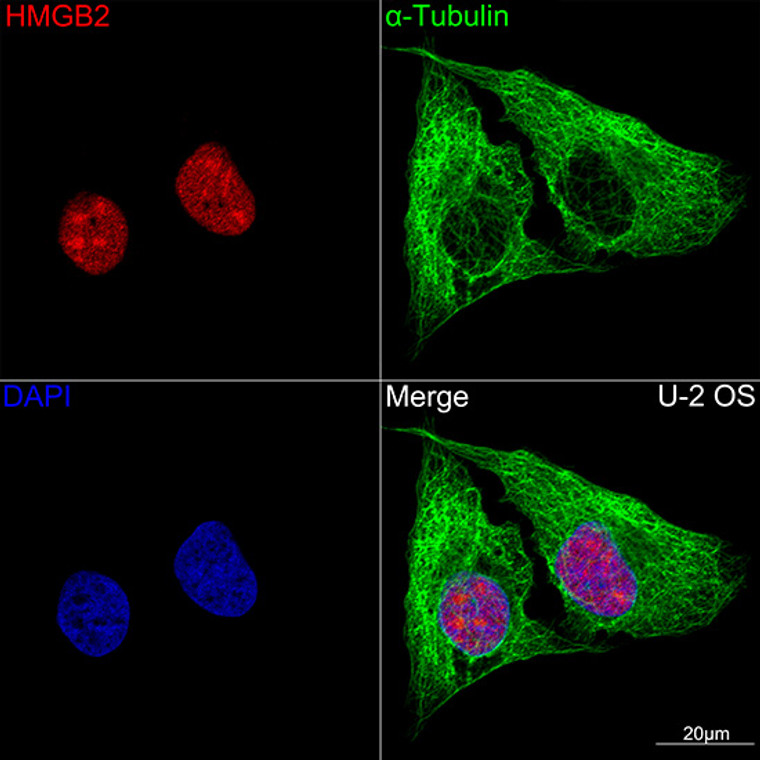
Rabbit monoclonal antibody anti-HMGB2 (1-100) is suitable for use in Western Blot, Immunohistochemistry, Immunofluorescence and Immunoprecipitation research applications. |
| Clonality: |
Monoclonal |
| Clone ID: |
S4MR |
| Conjugation: |
Unconjugated |
| Isotype: |
IgG |
| Formulation: |
PBS with 0.02% Sodium Azide, 0.05% BSA, 50% Glycerol, pH7.3. |
| Purification: |
Affinity purification |
| Dilution Range: |
WB 1:500-1:1000IHC-P 1:50-1:200IF/ICC 1:50-1:200IP 1:500-1:1000ChIP 1:500-1:1000CUT&Tag 10⁵ cells/1 Mu g |
| Storage Instruction: |
Store at-20°C for up to 1 year from the date of receipt, and avoid repeat freeze-thaw cycles. |
| Gene Symbol: |
HMGB2 |
| Gene ID: |
3148 |
| Uniprot ID: |
HMGB2_HUMAN |
| Immunogen Region: |
1-100 |
| Immunogen: |
A synthetic peptide corresponding to a sequence within amino acids 1-100 of human HMGB2 (P26583). |
| Immunogen Sequence: |
MGKGDPNKPRGKMSSYAFFV QTCREEHKKKHPDSSVNFAE FSKKCSERWKTMSAKEKSKF EDMAKSDKARYDREMKNYVP PKGDKKGKKKDPNAPKRPPS |
| Tissue Specificity | Expressed in gastric and intestinal tissues (at protein level). |
| Post Translational Modifications | Reduction/oxidation of cysteine residues Cys-23, Cys-45 and Cys-106 and a possible intramolecular disulfide bond involving Cys-23 and Cys-45 give rise to different redox forms with specific functional activities in various cellular compartments: 1- fully reduced HMGB2 (HMGB2C23hC45hC106h), 2- disulfide HMGB2 (HMGB2C23-C45C106h) and 3- sulfonyl HMGB2 (HMGB2C23soC45soC106so). Acetylation enhances nucleosome binding and chromation remodeling activity. |
| Function | Multifunctional protein with various roles in different cellular compartments. May act in a redox sensitive manner. In the nucleus is an abundant chromatin-associated non-histone protein involved in transcription, chromatin remodeling and V(D)J recombination and probably other processes. Binds DNA with a preference to non-canonical DNA structures such as single-stranded DNA. Can bent DNA and enhance DNA flexibility by looping thus providing a mechanism to promote activities on various gene promoters by enhancing transcription factor binding and/or bringing distant regulatory sequences into close proximity. Involved in V(D)J recombination by acting as a cofactor of the RAG complex: acts by stimulating cleavage and RAG protein binding at the 23 bp spacer of conserved recombination signal sequences (RSS). Proposed to be involved in the innate immune response to nucleic acids by acting as a promiscuous immunogenic DNA/RNA sensor which cooperates with subsequent discriminative sensing by specific pattern recognition receptors. In the extracellular compartment acts as a chemokine. Promotes proliferation and migration of endothelial cells implicating AGER/RAGE. Has antimicrobial activity in gastrointestinal epithelial tissues. Involved in inflammatory response to antigenic stimulus coupled with pro-inflammatory activity. Involved in modulation of neurogenesis probably by regulation of neural stem proliferation. Involved in articular cartilage surface maintenance implicating LEF1 and the Wnt/beta-catenin pathway. |
| Protein Name | High Mobility Group Protein B2High Mobility Group Protein 2Hmg-2 |
| Database Links | Reactome: R-HSA-140342 |
| Cellular Localisation | NucleusChromosomeCytoplasmSecretedIn Basal State Predominantly Nuclear |
| Alternative Antibody Names | Anti-High Mobility Group Protein B2 antibodyAnti-High Mobility Group Protein 2 antibodyAnti-Hmg-2 antibodyAnti-HMGB2 antibodyAnti-HMG2 antibody |
Information sourced from Uniprot.org
12 months for antibodies. 6 months for ELISA Kits. Please see website T&Cs for further guidance


