| Host: |
Rabbit |
| Applications: |
WB/IHC |
| Reactivity: |
Human/Mouse |
| Note: |
STRICTLY FOR FURTHER SCIENTIFIC RESEARCH USE ONLY (RUO). MUST NOT TO BE USED IN DIAGNOSTIC OR THERAPEUTIC APPLICATIONS. |
| Short Description: |
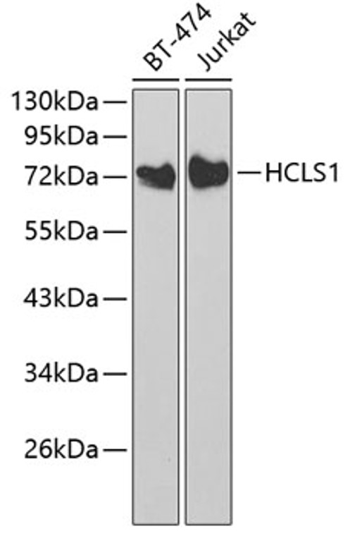
Rabbit polyclonal antibody anti-HCLS1 (1-280) is suitable for use in Western Blot and Immunohistochemistry research applications. |
| Clonality: |
Polyclonal |
| Conjugation: |
Unconjugated |
| Isotype: |
IgG |
| Formulation: |
PBS with 0.02% Sodium Azide, 50% Glycerol, pH7.3. |
| Purification: |
Affinity purification |
| Dilution Range: |
WB 1:500-1:2000IHC-P 1:50-1:200 |
| Storage Instruction: |
Store at-20°C for up to 1 year from the date of receipt, and avoid repeat freeze-thaw cycles. |
| Gene Symbol: |
HCLS1 |
| Gene ID: |
3059 |
| Uniprot ID: |
HCLS1_HUMAN |
| Immunogen Region: |
1-280 |
| Immunogen: |
Recombinant fusion protein containing a sequence corresponding to amino acids 1-280 of human HCLS1 (NP_005326.2). |
| Immunogen Sequence: |
MWKSVVGHDVSVSVETQGDD WDTDPDFVNDISEKEQRWGA KTIEGSGRTEHINIHQLRNK VSEEHDVLRKKEMESGPKAS HGYGGRFGVERDRMDKSAVG HEYVAEVEKHSSQTDAAKGF GGKYGVERDRADKSAVGFDY KGEVEKHTSQKDYSRGFGGR YGVEKDKWDKAALGYDYKGE TEKHESQRDYAKGFGGQYGI QKDRVDKSAVGFNEMEAPTT AYKKTTPIEAASSGTRGLK |
| Tissue Specificity | Expressed only in tissues and cells of hematopoietic origin. |
| Post Translational Modifications | Phosphorylated by FES. Phosphorylated by LYN, FYN and FGR after cross-linking of surface IgM on B-cells. Phosphorylation by LYN, FYN and FGR requires prior phosphorylation by SYK or FES. |
| Function | Substrate of the antigen receptor-coupled tyrosine kinase. Plays a role in antigen receptor signaling for both clonal expansion and deletion in lymphoid cells. May also be involved in the regulation of gene expression. |
| Protein Name | Hematopoietic Lineage Cell-Specific ProteinHematopoietic Cell-Specific Lyn Substrate 1Lckbp1P75 |
| Cellular Localisation | MembranePeripheral Membrane ProteinCytoplasmMitochondrion |
| Alternative Antibody Names | Anti-Hematopoietic Lineage Cell-Specific Protein antibodyAnti-Hematopoietic Cell-Specific Lyn Substrate 1 antibodyAnti-Lckbp1 antibodyAnti-P75 antibodyAnti-HCLS1 antibodyAnti-HS1 antibody |
Information sourced from Uniprot.org
12 months for antibodies. 6 months for ELISA Kits. Please see website T&Cs for further guidance


